5DWC | pdb_00005dwc

Crystal structure of restriction endonuclease AgeI


Experimental Data Snapshot

  • Method: X-RAY DIFFRACTION
  • Resolution: 2.50 Å
  • R-Value Free: 
    0.256 (Depositor), 0.259 (DCC) 
  • R-Value Work: 
    0.214 (Depositor), 0.222 (DCC) 
  • R-Value Observed: 
    0.219 (Depositor) 

Starting Model: experimental
View more details

wwPDB Validation 3D Report Full Report

Validation slider image for 5DWC

This is version 1.5 of the entry. See complete history

Literature

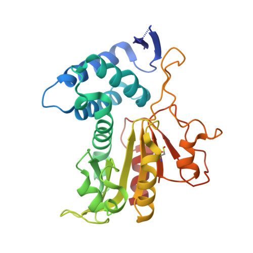
Restriction endonuclease AgeI is a monomer which dimerizes to cleave DNA.

Tamulaitiene, G.Jovaisaite, V.Tamulaitis, G.Songailiene, I.Manakova, E.Zaremba, M.Grazulis, S.Xu, S.Y.Siksnys, V.

(2017) Nucleic Acids Res 45: 3547-3558

  • DOI: https://doi.org/10.1093/nar/gkw1310
  • Primary Citation Related Structures: 
    5DWA, 5DWB, 5DWC

  • PubMed Abstract: 

    Although all Type II restriction endonucleases catalyze phosphodiester bond hydrolysis within or close to their DNA target sites, they form different oligomeric assemblies ranging from monomers, dimers, tetramers to higher order oligomers to generate a double strand break in DNA. Type IIP restriction endonuclease AgeI recognizes a palindromic sequence 5΄-A/CCGGT-3΄ and cuts it ('/' denotes the cleavage site) producing staggered DNA ends. Here, we present crystal structures of AgeI in apo and DNA-bound forms. The structure of AgeI is similar to the restriction enzymes that share in their target sites a conserved CCGG tetranucleotide and a cleavage pattern. Structure analysis and biochemical data indicate, that AgeI is a monomer in the apo-form both in the crystal and in solution, however, it binds and cleaves the palindromic target site as a dimer. DNA cleavage mechanism of AgeI is novel among Type IIP restriction endonucleases.


  • Organizational Affiliation
    • Institute of Biotechnology, Vilnius University, Sauletekio al. 7, LT-10257 Vilnius, Lithuania.

Macromolecule Content 

  • Total Structure Weight: 30.59 kDa 
  • Atom Count: 2,145 
  • Modeled Residue Count: 274 
  • Deposited Residue Count: 278 
  • Unique protein chains: 1

Macromolecules

Find similar proteins by:|  3D Structure
Entity ID: 1
MoleculeChains  Sequence LengthOrganismDetailsImage
Type-2 restriction enzyme AgeI278Thalassovita gelatinovoraMutation(s): 0 
Gene Names: ageIR
EC: 3.1.21.4
UniProt
Find proteins for Q9KHV6 (Thalassovita gelatinovora)
Explore Q9KHV6 
Go to UniProtKB:  Q9KHV6
Entity Groups
Sequence Clusters30% Identity50% Identity70% Identity90% Identity95% Identity100% Identity
UniProt GroupQ9KHV6
Sequence Annotations
Expand
Reference Sequence

Small Molecules

Modified Residues  1 Unique
IDChains TypeFormula2D DiagramParent
CSO
Query on CSO
A
L-PEPTIDE LINKINGC3 H7 N O3 SCYS

Experimental Data & Validation

Experimental Data

  • Method: X-RAY DIFFRACTION
  • Resolution: 2.50 Å
  • R-Value Free:  0.256 (Depositor), 0.259 (DCC) 
  • R-Value Work:  0.214 (Depositor), 0.222 (DCC) 
  • R-Value Observed: 0.219 (Depositor) 
Space Group: P 21 21 21
Unit Cell:
Length ( Å )Angle ( ˚ )
a = 37.213α = 90
b = 59.269β = 90
c = 120.915γ = 90
Software Package:
Software NamePurpose
MxCuBEdata collection
MOSFLMdata reduction
SCALAdata scaling
MOLREPphasing
Cootmodel building
PHENIXrefinement
PDB_EXTRACTdata extraction

Structure Validation

View Full Validation Report


Entry History 

& Funding Information

Deposition Data


Funding OrganizationLocationGrant Number
Research Council of LithuaniaLithuaniaMIP-41/2013

Revision History  (Full details and data files)

  • Version 1.0: 2016-09-28
    Type: Initial release
  • Version 1.1: 2017-01-11
    Changes: Database references
  • Version 1.2: 2017-05-03
    Changes: Database references
  • Version 1.3: 2018-01-17
    Changes: Data collection
  • Version 1.4: 2024-01-10
    Changes: Data collection, Database references, Refinement description
  • Version 1.5: 2024-11-13
    Changes: Structure summary