5DTQ | pdb_00005dtq

Crystal structure of Dot1L in complex with inhibitor CPD3 [(2,6-dichlorophenyl)(quinolin-6-yl)methanone]


Experimental Data Snapshot

  • Method: X-RAY DIFFRACTION
  • Resolution: 2.61 Å
  • R-Value Free: 
    0.208 (Depositor), 0.204 (DCC) 
  • R-Value Work: 
    0.173 (Depositor), 0.173 (DCC) 
  • R-Value Observed: 
    0.175 (Depositor) 

Starting Model: experimental
View more details

wwPDB Validation 3D Report Full Report

Validation slider image for 5DTQ

Ligand Structure Quality Assessment 


This is version 1.2 of the entry. See complete history

Literature

Optimization of a Fragment-Based Screening Hit toward Potent DOT1L Inhibitors Interacting in an Induced Binding Pocket.

Scheufler, C.Mobitz, H.Gaul, C.Ragot, C.Be, C.Fernandez, C.Beyer, K.S.Tiedt, R.Stauffer, F.

(2016) ACS Med Chem Lett 7: 730-734

  • DOI: https://doi.org/10.1021/acsmedchemlett.6b00168
  • Primary Citation Related Structures: 
    5DTM, 5DTQ, 5DTR

  • PubMed Abstract: 

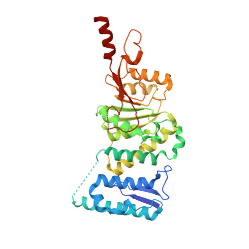
    Mixed lineage leukemia (MLL) gene rearrangement induces leukemic transformation by ectopic recruitment of disruptor of telomeric silencing 1-like protein (DOT1L), a lysine histone methyltransferase, leading to local hypermethylation of H3K79 and misexpression of genes (including HoxA), which drive the leukemic phenotype. A weak fragment-based screening hit identified by SPR was cocrystallized with DOT1L and optimized using structure-based ligand optimization to yield compound 8 (IC50 = 14 nM). This series of inhibitors is structurally not related to cofactor SAM and is not interacting within the SAM binding pocket but induces a pocket adjacent to the SAM binding site.


  • Organizational Affiliation
    • Novartis Institutes for Biomedical Research , 4002 Basel, Switzerland.

Macromolecule Content 

  • Total Structure Weight: 77.52 kDa 
  • Atom Count: 5,389 
  • Modeled Residue Count: 639 
  • Deposited Residue Count: 668 
  • Unique protein chains: 1

Macromolecules

Find similar proteins by:|  3D Structure
Entity ID: 1
MoleculeChains  Sequence LengthOrganismDetailsImage
Histone-lysine N-methyltransferase, H3 lysine-79 specific
A, B
334Homo sapiensMutation(s): 0 
Gene Names: DOT1LKIAA1814KMT4
EC: 2.1.1.43 (PDB Primary Data), 2.1.1.360 (UniProt)
UniProt & NIH Common Fund Data Resources
Find proteins for Q8TEK3 (Homo sapiens)
Explore Q8TEK3 
Go to UniProtKB:  Q8TEK3
PHAROS:  Q8TEK3
GTEx:  ENSG00000104885 
Entity Groups
Sequence Clusters30% Identity50% Identity70% Identity90% Identity95% Identity100% Identity
UniProt GroupQ8TEK3
Sequence Annotations
Expand
Reference Sequence

Small Molecules

Binding Affinity Annotations 
IDSourceBinding Affinity
5F6 BindingDB:  5DTQ IC50: 1.39e+5 (nM) from 1 assay(s)

Experimental Data & Validation

Experimental Data

  • Method: X-RAY DIFFRACTION
  • Resolution: 2.61 Å
  • R-Value Free:  0.208 (Depositor), 0.204 (DCC) 
  • R-Value Work:  0.173 (Depositor), 0.173 (DCC) 
  • R-Value Observed: 0.175 (Depositor) 
Space Group: P 63
Unit Cell:
Length ( Å )Angle ( ˚ )
a = 158.651α = 90
b = 158.651β = 90
c = 74.179γ = 120
Software Package:
Software NamePurpose
BUSTERrefinement
XDSdata reduction
XSCALEdata scaling
PHASERphasing

Structure Validation

View Full Validation Report


Ligand Structure Quality Assessment 


Entry History 

Deposition Data

Revision History  (Full details and data files)

  • Version 1.0: 2016-06-15
    Type: Initial release
  • Version 1.1: 2016-09-07
    Changes: Database references
  • Version 1.2: 2024-01-10
    Changes: Data collection, Database references, Refinement description