5DQO

Crystal structure of Y347F mutant of human primase p58 iron-sulfur cluster domain


Experimental Data Snapshot

  • Method: X-RAY DIFFRACTION
  • Resolution: 2.30 Å
  • R-Value Free: 0.275 
  • R-Value Work: 0.251 
  • R-Value Observed: 0.252 

wwPDB Validation   3D Report Full Report


This is version 1.5 of the entry. See complete history


Literature

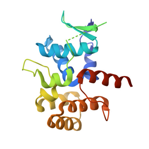
The [4Fe4S] cluster of human DNA primase functions as a redox switch using DNA charge transport.

O'Brien, E.Holt, M.E.Thompson, M.K.Salay, L.E.Ehlinger, A.C.Chazin, W.J.Barton, J.K.

(2017) Science 355

  • DOI: https://doi.org/10.1126/science.aag1789
  • Primary Citation of Related Structures:  
    5DQO, 5I7M

  • PubMed Abstract: 

    DNA charge transport chemistry offers a means of long-range, rapid redox signaling. We demonstrate that the [4Fe4S] cluster in human DNA primase can make use of this chemistry to coordinate the first steps of DNA synthesis. Using DNA electrochemistry, we found that a change in oxidation state of the [4Fe4S] cluster acts as a switch for DNA binding. Single-atom mutations that inhibit this charge transfer hinder primase initiation without affecting primase structure or polymerization. Generating a single base mismatch in the growing primer duplex, which attenuates DNA charge transport, inhibits primer truncation. Thus, redox signaling by [4Fe4S] clusters using DNA charge transport regulates primase binding to DNA and illustrates chemistry that may efficiently drive substrate handoff between polymerases during DNA replication.


  • Organizational Affiliation

    Division of Chemistry and Chemical Engineering, California Institute of Technology, Pasadena, CA 91125, USA.


Macromolecules
Find similar proteins by:  (by identity cutoff)  |  3D Structure
Entity ID: 1
MoleculeChains Sequence LengthOrganismDetailsImage
DNA primase large subunit
A, B, C, D
197Homo sapiensMutation(s): 1 
Gene Names: PRIM2PRIM2A
EC: 2.7.7
UniProt & NIH Common Fund Data Resources
Find proteins for P49643 (Homo sapiens)
Explore P49643 
Go to UniProtKB:  P49643
PHAROS:  P49643
GTEx:  ENSG00000146143 
Entity Groups  
Sequence Clusters30% Identity50% Identity70% Identity90% Identity95% Identity100% Identity
UniProt GroupP49643
Sequence Annotations
Expand
  • Reference Sequence
Experimental Data & Validation

Experimental Data

  • Method: X-RAY DIFFRACTION
  • Resolution: 2.30 Å
  • R-Value Free: 0.275 
  • R-Value Work: 0.251 
  • R-Value Observed: 0.252 
  • Space Group: P 31
Unit Cell:
Length ( Å )Angle ( ˚ )
a = 60.398α = 90
b = 60.398β = 90
c = 246.729γ = 120
Software Package:
Software NamePurpose
REFMACrefinement
HKL-2000data collection
SCALEPACKdata scaling
PHASERphasing
PDB_EXTRACTdata extraction
HKL-2000data reduction

Structure Validation

View Full Validation Report


Entry History & Funding Information

Deposition Data


Funding OrganizationLocationGrant Number
National Institutes of Health/National Institute of General Medical Sciences (NIH/NIGMS)United StatesGM65484

Revision History  (Full details and data files)

  • Version 1.0: 2016-09-21
    Type: Initial release
  • Version 1.1: 2016-10-05
    Changes: Database references
  • Version 1.2: 2017-03-08
    Changes: Database references
  • Version 1.3: 2017-09-13
    Changes: Author supporting evidence
  • Version 1.4: 2019-12-25
    Changes: Author supporting evidence
  • Version 1.5: 2023-09-27
    Changes: Data collection, Database references, Refinement description