


Crystal structure of UbiG mutant in complex with SAH at 2.1 angstroms resolution
Zhu, Y., Jiang, X., Li, X., Teng, M.To be published.
Experimental Data Snapshot
Starting Model: experimental
View more details
Entity ID: 1 | |||||
|---|---|---|---|---|---|
| Molecule | Chains | Sequence Length | Organism | Details | Image |
| Ubiquinone biosynthesis O-methyltransferase | 225 | Escherichia coli HS | Mutation(s): 0 Gene Names: ubiG, EcHS_A2372 EC: 2.1.1.222 (PDB Primary Data), 2.1.1.64 (PDB Primary Data) | 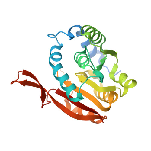 | |
UniProt | |||||
Entity Groups | |||||
| Sequence Clusters | 30% Identity50% Identity70% Identity90% Identity95% Identity100% Identity | ||||
| UniProt Group | P17993 | ||||
Sequence AnnotationsExpand | |||||
Reference Sequence | |||||
| Ligands 1 Unique | |||||
|---|---|---|---|---|---|
| ID | Chains | Name / Formula / InChI Key | 2D Diagram | 3D Interactions | |
| SAH Download:Ideal Coordinates CCD File | B [auth A] | S-ADENOSYL-L-HOMOCYSTEINE C14 H20 N6 O5 S ZJUKTBDSGOFHSH-WFMPWKQPSA-N |  | ||
| Length ( Å ) | Angle ( ˚ ) |
|---|---|
| a = 139.842 | α = 90 |
| b = 39.326 | β = 94.34 |
| c = 40.112 | γ = 90 |
| Software Name | Purpose |
|---|---|
| PHENIX | refinement |
| HKL-2000 | data reduction |
| SCALA | data scaling |
| MOLREP | phasing |


