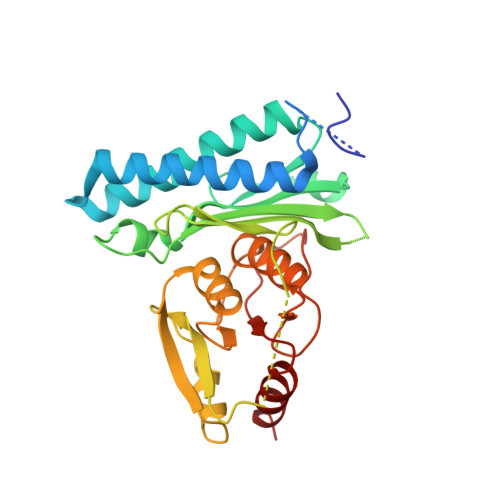
Crystal Structures of Mycobacterium tuberculosis CysQ, with Substrate and Products Bound.
Erickson, A.I., Sarsam, R.D., Fisher, A.J.(2015) Biochemistry 54: 6830-6841
- PubMed: 26512869 Search on PubMed
- DOI: https://doi.org/10.1021/acs.biochem.5b01000
- Primary Citation Related Structures:
5DJF, 5DJG, 5DJH, 5DJI, 5DJJ, 5DJK - PubMed Abstract:
In many organisms, 3'-phosphoadenosine 5'-phosphate (PAP) is a product of two reactions in the sulfur activation pathway. The sulfurylation of biomolecules, catalyzed by sulfotransferases, uses 3'-phosphoadenosine 5'-phosphosulfate (PAPS) as a sulfate donor, producing the sulfated biomolecule and PAP product. Additionally, the first step in sulfate reduction for many bacteria and fungi reduces the sulfate moiety of PAPS, producing PAP and sulfite, which is subsequently reduced to sulfide. PAP is removed by the phosphatase activity of CysQ, a 3',5'-bisphosphate nucleotidase, yielding AMP and phosphate. Because excess PAP alters the equilibrium of the sulfur pathway and inhibits sulfotransferases, PAP concentrations can affect the levels of sulfur-containing metabolites. Therefore, CysQ, a divalent cation metal-dependent phosphatase, is a major regulator of this pathway. CysQ (Rv2131c) from Mycobacterium tuberculosis (Mtb) was successfully expressed, purified, and crystallized in a variety of ligand-bound states. Here we report six crystal structures of Mtb CysQ, including a ligand-free structure, a lithium-inhibited state with substrate PAP bound, and a product-bound complex with AMP, phosphate, and three Mg(2+) ions bound. Comparison of these structures together with homologues of the superfamily has provided insight into substrate specificity, metal coordination, and catalytic mechanism.
- Department of Chemistry, ‡Department of Molecular and Cellular Biology, and §Graduate Program in Biochemistry and Molecular, Cellular and Developmental Biology, University of California , One Shields Avenue, Davis, California 95616, United States.
Organizational Affiliation: