5DGO | pdb_00005dgo

Crystal structure of cell division cycle protein 45 (Cdc45)


Experimental Data Snapshot

  • Method: X-RAY DIFFRACTION
  • Resolution: 2.10 Å
  • R-Value Free: 
    0.195 (Depositor), 0.197 (DCC) 
  • R-Value Work: 
    0.168 (Depositor), 0.171 (DCC) 
  • R-Value Observed: 
    0.169 (Depositor) 

wwPDB Validation 3D Report Full Report

Validation slider image for 5DGO

This is version 1.1 of the entry. See complete history

Literature

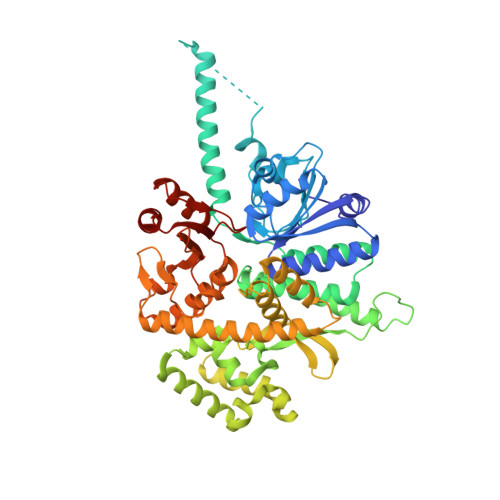
Structure of human Cdc45 and implications for CMG helicase function.

Simon, A.C.Sannino, V.Costanzo, V.Pellegrini, L.

(2016) Nat Commun 7: 11638-11638

  • DOI: https://doi.org/10.1038/ncomms11638
  • Primary Citation Related Structures: 
    5DGO

  • PubMed Abstract: 

    Cell division cycle protein 45 (Cdc45) is required for DNA synthesis during genome duplication, as a component of the Cdc45-MCM-GINS (CMG) helicase. Despite its essential biological function, its biochemical role in DNA replication has remained elusive. Here we report the 2.1-Å crystal structure of human Cdc45, which confirms its evolutionary link with the bacterial RecJ nuclease and reveals several unexpected features that underpin its function in eukaryotic DNA replication. These include a long-range interaction between N- and C-terminal DHH domains, blocking access to the DNA-binding groove of its RecJ-like fold, and a helical insertion in its N-terminal DHH domain, which appears poised for replisome interactions. In combination with available electron microscopy data, we validate by mutational analysis the mechanism of Cdc45 association with the MCM ring and GINS co-activator, critical for CMG assembly. These findings provide an indispensable molecular basis to rationalize the essential role of Cdc45 in genomic duplication.


  • Organizational Affiliation
    • Department of Biochemistry, University of Cambridge, Cambridge CB2 1GA, UK.

Macromolecule Content 

  • Total Structure Weight: 64.94 kDa 
  • Atom Count: 5,193 
  • Modeled Residue Count: 541 
  • Deposited Residue Count: 555 
  • Unique protein chains: 1

Macromolecules

Find similar proteins by:|  3D Structure
Entity ID: 1
MoleculeChains  Sequence LengthOrganismDetailsImage
Cell division control protein 45 homolog555Homo sapiensMutation(s): 0 
Gene Names: CDC45CDC45LCDC45L2UNQ374/PRO710
UniProt & NIH Common Fund Data Resources
Find proteins for O75419 (Homo sapiens)
Explore O75419 
Go to UniProtKB:  O75419
PHAROS:  O75419
GTEx:  ENSG00000093009 
Entity Groups
Sequence Clusters30% Identity50% Identity70% Identity90% Identity95% Identity100% Identity
UniProt GroupO75419
Sequence Annotations
Expand
Reference Sequence

Small Molecules

Ligands 2 Unique
IDChains Name / Formula / InChI Key2D Diagram3D Interactions
SO4

Query on SO4


Download:Ideal Coordinates CCD File
H [auth A],
I [auth A],
J [auth A]
SULFATE ION
O4 S
QAOWNCQODCNURD-UHFFFAOYSA-L
EDO

Query on EDO


Download:Ideal Coordinates CCD File
B [auth A]
C [auth A]
D [auth A]
E [auth A]
F [auth A]
B [auth A],
C [auth A],
D [auth A],
E [auth A],
F [auth A],
G [auth A]
1,2-ETHANEDIOL
C2 H6 O2
LYCAIKOWRPUZTN-UHFFFAOYSA-N

Experimental Data & Validation

Experimental Data

  • Method: X-RAY DIFFRACTION
  • Resolution: 2.10 Å
  • R-Value Free:  0.195 (Depositor), 0.197 (DCC) 
  • R-Value Work:  0.168 (Depositor), 0.171 (DCC) 
  • R-Value Observed: 0.169 (Depositor) 
Space Group: P 31 2 1
Unit Cell:
Length ( Å )Angle ( ˚ )
a = 112.795α = 90
b = 112.795β = 90
c = 143.06γ = 120
Software Package:
Software NamePurpose
PHENIXrefinement
XDSdata reduction
Aimlessdata scaling
PHENIXphasing

Structure Validation

View Full Validation Report


Entry History 

& Funding Information

Deposition Data


Funding OrganizationLocationGrant Number
Wellcome TrustUnited Kingdom104641/Z/14/Z

Revision History  (Full details and data files)

  • Version 1.0: 2016-05-25
    Type: Initial release
  • Version 1.1: 2024-11-13
    Changes: Data collection, Database references, Structure summary