Altiratinib Inhibits Tumor Growth, Invasion, Angiogenesis, and Microenvironment-Mediated Drug Resistance via Balanced Inhibition of MET, TIE2, and VEGFR2.
Smith, B.D., Kaufman, M.D., Leary, C.B., Turner, B.A., Wise, S.C., Ahn, Y.M., Booth, R.J., Caldwell, T.M., Ensinger, C.L., Hood, M.M., Lu, W.P., Patt, T.W., Patt, W.C., Rutkoski, T.J., Samarakoon, T., Telikepalli, H., Vogeti, L., Vogeti, S., Yates, K.M., Chun, L., Stewart, L.J., Clare, M., Flynn, D.L.(2015) Mol Cancer Ther 14: 2023-2034
- PubMed: 26285778 Search on PubMed
- DOI: https://doi.org/10.1158/1535-7163.MCT-14-1105
- Primary Citation Related Structures:
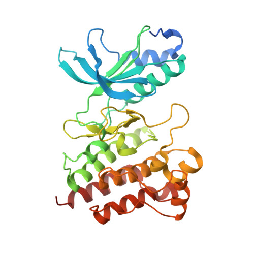
5DG5 - PubMed Abstract:
Altiratinib (DCC-2701) was designed based on the rationale of engineering a single therapeutic agent able to address multiple hallmarks of cancer (1). Specifically, altiratinib inhibits not only mechanisms of tumor initiation and progression, but also drug resistance mechanisms in the tumor and microenvironment through balanced inhibition of MET, TIE2 (TEK), and VEGFR2 (KDR) kinases. This profile was achieved by optimizing binding into the switch control pocket of all three kinases, inducing type II inactive conformations. Altiratinib durably inhibits MET, both wild-type and mutated forms, in vitro and in vivo. Through its balanced inhibitory potency versus MET, TIE2, and VEGFR2, altiratinib provides an agent that inhibits three major evasive (re)vascularization and resistance pathways (HGF, ANG, and VEGF) and blocks tumor invasion and metastasis. Altiratinib exhibits properties amenable to oral administration and exhibits substantial blood-brain barrier penetration, an attribute of significance for eventual treatment of brain cancers and brain metastases.
- Deciphera Pharmaceuticals, Lawrence, Kansas.
Organizational Affiliation: