5DEY | pdb_00005dey

Crystal structure of PAK1 in complex with an inhibitor compound G-5555


Experimental Data Snapshot

  • Method: X-RAY DIFFRACTION
  • Resolution: 2.10 Å
  • R-Value Free: 
    0.228 (Depositor), 0.228 (DCC) 
  • R-Value Work: 
    0.183 (Depositor), 0.187 (DCC) 
  • R-Value Observed: 
    0.186 (Depositor) 

wwPDB Validation 3D Report Full Report

Validation slider image for 5DEY

Ligand Structure Quality Assessment 


This is version 1.2 of the entry. See complete history

Literature

Design of Selective PAK1 Inhibitor G-5555: Improving Properties by Employing an Unorthodox Low-pK a Polar Moiety.

Ndubaku, C.O.Crawford, J.J.Drobnick, J.Aliagas, I.Campbell, D.Dong, P.Dornan, L.M.Duron, S.Epler, J.Gazzard, L.Heise, C.E.Hoeflich, K.P.Jakubiak, D.La, H.Lee, W.Lin, B.Lyssikatos, J.P.Maksimoska, J.Marmorstein, R.Murray, L.J.O'Brien, T.Oh, A.Ramaswamy, S.Wang, W.Zhao, X.Zhong, Y.Blackwood, E.Rudolph, J.

(2015) ACS Med Chem Lett 6: 1241-1246

  • DOI: https://doi.org/10.1021/acsmedchemlett.5b00398
  • Primary Citation Related Structures: 
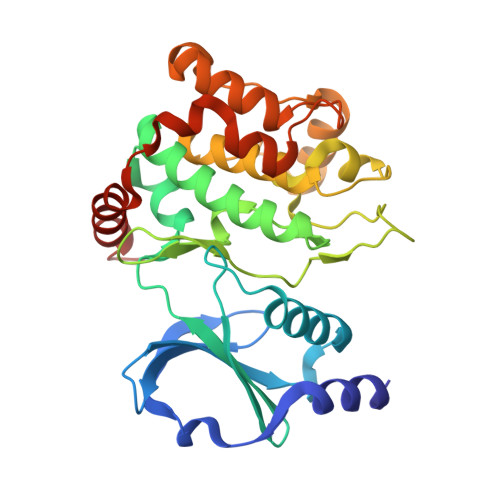
    5DEY, 5DFP

  • PubMed Abstract: 

    Signaling pathways intersecting with the p21-activated kinases (PAKs) play important roles in tumorigenesis and cancer progression. By recognizing that the limitations of FRAX1036 (1) were chiefly associated with the highly basic amine it contained, we devised a mitigation strategy to address several issues such as hERG activity. The 5-amino-1,3-dioxanyl moiety was identified as an effective means of reducing pK a and logP simultaneously. When positioned properly within the scaffold, this group conferred several benefits including potency, pharmacokinetics, and selectivity. Mouse xenograft PK/PD studies were carried out using an advanced compound, G-5555 (12), derived from this approach. These studies concluded that dose-dependent pathway modulation was achievable and paves the way for further in vivo investigations of PAK1 function in cancer and other diseases.


  • Organizational Affiliation
    • Genentech, Inc. , 1 DNA Way, South San Francisco, California 94080, United States.

Macromolecule Content 

  • Total Structure Weight: 67.56 kDa 
  • Atom Count: 4,762 
  • Modeled Residue Count: 576 
  • Deposited Residue Count: 594 
  • Unique protein chains: 1

Macromolecules

Find similar proteins by:|  3D Structure
Entity ID: 1
MoleculeChains  Sequence LengthOrganismDetailsImage
Serine/threonine-protein kinase PAK 1
A, B
297Homo sapiensMutation(s): 2 
Gene Names: PAK1
EC: 2.7.11.1
UniProt & NIH Common Fund Data Resources
Find proteins for Q13153 (Homo sapiens)
Explore Q13153 
Go to UniProtKB:  Q13153
PHAROS:  Q13153
GTEx:  ENSG00000149269 
Entity Groups
Sequence Clusters30% Identity50% Identity70% Identity90% Identity95% Identity100% Identity
UniProt GroupQ13153
Sequence Annotations
Expand
Reference Sequence

Small Molecules

Ligands 1 Unique
IDChains Name / Formula / InChI Key2D Diagram3D Interactions
59T

Query on 59T


Download:Ideal Coordinates CCD File
C [auth A],
D [auth B]
8-[(trans-5-amino-1,3-dioxan-2-yl)methyl]-6-[2-chloro-4-(6-methylpyridin-2-yl)phenyl]-2-(methylamino)pyrido[2,3-d]pyrimidin-7(8H)-one
C25 H25 Cl N6 O3
ZBCMHWUFWQFPLV-VVOJOOEHSA-N

Experimental Data & Validation

Experimental Data

  • Method: X-RAY DIFFRACTION
  • Resolution: 2.10 Å
  • R-Value Free:  0.228 (Depositor), 0.228 (DCC) 
  • R-Value Work:  0.183 (Depositor), 0.187 (DCC) 
  • R-Value Observed: 0.186 (Depositor) 
Space Group: P 1 21 1
Unit Cell:
Length ( Å )Angle ( ˚ )
a = 61.661α = 90
b = 81.863β = 106.4
c = 65.916γ = 90
Software Package:
Software NamePurpose
PHENIXrefinement
HKL-2000data reduction
SCALEPACKdata scaling
PHASERphasing

Structure Validation

View Full Validation Report


Ligand Structure Quality Assessment 


Entry History 

Deposition Data

Revision History  (Full details and data files)

  • Version 1.0: 2016-01-27
    Type: Initial release
  • Version 1.1: 2016-06-01
    Changes: Data collection
  • Version 1.2: 2024-10-16
    Changes: Data collection, Database references, Derived calculations, Structure summary