5DDO | pdb_00005ddo

Structural and Dynamic Basis for Low Affinity-High Selectivity Binding of L-glutamine by the Gln-riboswitch


Experimental Data Snapshot

  • Method: X-RAY DIFFRACTION
  • Resolution: 3.10 Å
  • R-Value Free: 
    0.289 (Depositor), 0.285 (DCC) 
  • R-Value Work: 
    0.227 (Depositor), 0.226 (DCC) 
  • R-Value Observed: 
    0.231 (Depositor) 

Starting Model: experimental
View more details

wwPDB Validation 3D Report Full Report

Validation slider image for 5DDO

This is version 1.5 of the entry. See complete history

Literature

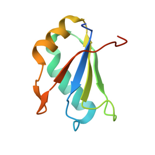
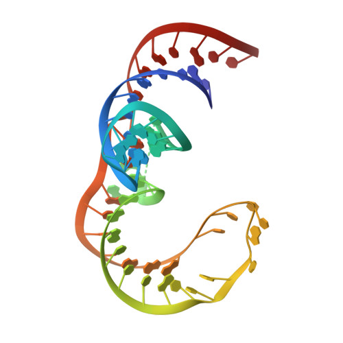
Structural and Dynamic Basis for Low-Affinity, High-Selectivity Binding of L-Glutamine by the Glutamine Riboswitch.

Ren, A.Xue, Y.Peselis, A.Serganov, A.Al-Hashimi, H.M.Patel, D.J.

(2015) Cell Rep 13: 1800-1813

  • DOI: https://doi.org/10.1016/j.celrep.2015.10.062
  • Primary Citation Related Structures: 
    5DDO, 5DDP, 5DDQ, 5DDR

  • PubMed Abstract: 

    Naturally occurring L-glutamine riboswitches occur in cyanobacteria and marine metagenomes, where they reside upstream of genes involved in nitrogen metabolism. By combining X-ray, NMR, and MD, we characterized an L-glutamine-dependent conformational transition in the Synechococcus elongatus glutamine riboswitch from tuning fork to L-shaped alignment of stem segments. This transition generates an open ligand-binding pocket with L-glutamine selectivity enforced by Mg(2+)-mediated intermolecular interactions. The transition also stabilizes the P1 helix through a long-range "linchpin" Watson-Crick G-C pair-capping interaction, while melting a short helix below P1 potentially capable of modulating downstream readout. NMR data establish that the ligand-free glutamine riboswitch in Mg(2+) solution exists in a slow equilibrium between flexible tuning fork and a minor conformation, similar, but not identical, to the L-shaped bound conformation. We propose that an open ligand-binding pocket combined with a high conformational penalty for forming the ligand-bound state provide mechanisms for reducing binding affinity while retaining high selectivity.


  • Organizational Affiliation
    • Structural Biology Program, Memorial Sloan-Kettering Cancer Center, New York, NY 10065, USA.

Macromolecule Content 

  • Total Structure Weight: 61.86 kDa 
  • Atom Count: 3,791 
  • Modeled Residue Count: 289 
  • Deposited Residue Count: 316 
  • Unique protein chains: 1
  • Unique nucleic acid chains: 1

Macromolecules


Find similar proteins by:|  3D Structure
Entity ID: 2
MoleculeChains  Sequence LengthOrganismDetailsImage
U1 small nuclear ribonucleoprotein AB [auth G],
D [auth C]
97Homo sapiensMutation(s): 2 
Gene Names: SNRPA
UniProt & NIH Common Fund Data Resources
Find proteins for P09012 (Homo sapiens)
Explore P09012 
Go to UniProtKB:  P09012
PHAROS:  P09012
GTEx:  ENSG00000077312 
Entity Groups
Sequence Clusters30% Identity50% Identity70% Identity90% Identity95% Identity100% Identity
UniProt GroupP09012
Sequence Annotations
Expand
Reference Sequence
Find similar nucleic acids by:  (by identity cutoff) 
Entity ID: 1
MoleculeChains LengthOrganismImage
L-glutamine riboswitch (58-MER)A,
C [auth B]
61Synechococcus elongatus
Sequence Annotations
Expand
Reference Sequence

Experimental Data & Validation

Experimental Data

  • Method: X-RAY DIFFRACTION
  • Resolution: 3.10 Å
  • R-Value Free:  0.289 (Depositor), 0.285 (DCC) 
  • R-Value Work:  0.227 (Depositor), 0.226 (DCC) 
  • R-Value Observed: 0.231 (Depositor) 
Space Group: C 1 2 1
Unit Cell:
Length ( Å )Angle ( ˚ )
a = 80.84α = 90
b = 99.652β = 99.05
c = 88.461γ = 90
Software Package:
Software NamePurpose
PHENIXrefinement
HKL-2000data reduction
HKL-2000data scaling
PHASERphasing

Structure Validation

View Full Validation Report


Entry History 

& Funding Information

Deposition Data


Funding OrganizationLocationGrant Number
National Institutes of Health/National Cancer Institute (NIH/NCI)United States1 U19 CA179564

Revision History  (Full details and data files)

  • Version 1.0: 2015-12-23
    Type: Initial release
  • Version 1.1: 2017-04-05
    Changes: Derived calculations
  • Version 1.2: 2017-09-20
    Changes: Author supporting evidence
  • Version 1.3: 2019-12-04
    Changes: Author supporting evidence
  • Version 1.4: 2024-03-06
    Changes: Data collection, Database references
  • Version 1.5: 2026-03-04
    Changes: Refinement description, Structure summary