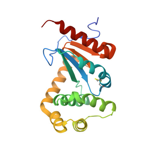
Fragment library screening identifies hits that bind to the non-catalytic surface of Pseudomonas aeruginosa DsbA1.
Mohanty, B., Rimmer, K., McMahon, R.M., Headey, S.J., Vazirani, M., Shouldice, S.R., Coincon, M., Tay, S., Morton, C.J., Simpson, J.S., Martin, J.L., Scanlon, M.J.(2017) PLoS One 12: e0173436-e0173436
- PubMed: 28346540 Search on PubMedSearch on PubMed Central
- DOI: https://doi.org/10.1371/journal.pone.0173436
- Primary Citation Related Structures:
2MBT, 5DCH, 5TLQ - PubMed Abstract:
At a time when the antibiotic drug discovery pipeline has stalled, antibiotic resistance is accelerating with catastrophic implications for our ability to treat bacterial infections. Globally we face the prospect of a future when common infections can once again kill. Anti-virulence approaches that target the capacity of the bacterium to cause disease rather than the growth or survival of the bacterium itself offer a tantalizing prospect of novel antimicrobials. They may also reduce the propensity to induce resistance by removing the strong selection pressure imparted by bactericidal or bacteriostatic agents. In the human pathogen Pseudomonas aeruginosa, disulfide bond protein A (PaDsbA1) plays a central role in the oxidative folding of virulence factors and is therefore an attractive target for the development of new anti-virulence antimicrobials. Using a fragment-based approach we have identified small molecules that bind to PaDsbA1. The fragment hits show selective binding to PaDsbA1 over the DsbA protein from Escherichia coli, suggesting that developing species-specific narrow-spectrum inhibitors of DsbA enzymes may be feasible. Structures of a co-complex of PaDsbA1 with the highest affinity fragment identified in the screen reveal that the fragment binds on the non-catalytic surface of the protein at a domain interface. This biophysical and structural data represent a starting point in the development of higher affinity compounds, which will be assessed for their potential as selective PaDsbA1 inhibitors.
- Medicinal Chemistry, Monash Institute of Pharmaceutical Sciences, Monash University, Parkville, Victoria, Australia.
Organizational Affiliation: