5D55 | pdb_00005d55

Crystal structure of the E. coli Hda pilus minor tip subunit, HdaB


Experimental Data Snapshot

  • Method: X-RAY DIFFRACTION
  • Resolution: 2.00 Å
  • R-Value Free: 
    0.248 (Depositor), 0.257 (DCC) 
  • R-Value Work: 
    0.203 (Depositor), 0.218 (DCC) 
  • R-Value Observed: 
    0.207 (Depositor) 

Starting Model: experimental
View more details

wwPDB Validation 3D Report Full Report

Validation slider image for 5D55

This is version 1.3 of the entry. See complete history

Literature

Crystal structure and analysis of HdaB: The enteroaggregative Escherichia coli AAF/IV pilus tip protein.

Lee, W.C.Matthews, S.Garnett, J.A.

(2016) Protein Sci 25: 1898-1905

  • DOI: https://doi.org/10.1002/pro.2982
  • Primary Citation Related Structures: 
    5D55

  • PubMed Abstract: 

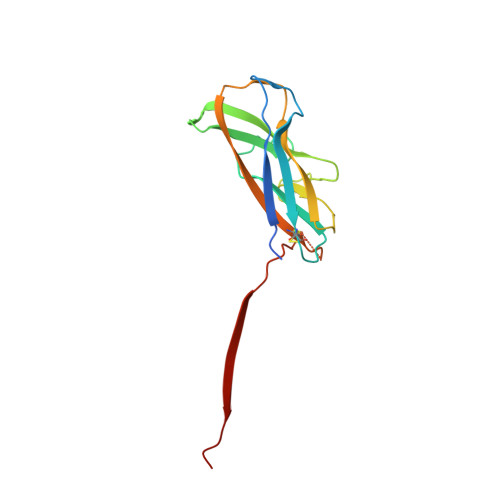
    Enteroaggregative Escherichia coli is the primary cause of pediatric diarrhea in developing countries. They utilize aggregative adherence fimbriae (AAFs) to promote initial adherence to the host intestinal mucosa, promote the formation of biofilms, and mediate host invasion. Five AAFs have been identified to date and AAF/IV is amongst the most prevalent found in clinical isolates. Here we present the X-ray crystal structure of the AAF/IV tip protein HdaB at 2.0 Å resolution. It shares high structural homology with members of the Afa/Dr superfamily of fimbriae, which are involved in host invasion. We highlight surface exposed residues that share sequence homology and propose that these may function in invasion and also non-conserved regions that could mediate HdaB specific adhesive functions.


  • Organizational Affiliation
    • Department of Life Sciences, Centre for Structural Biology, Imperial College London, South Kensington, London, SW7 2AZ, United Kingdom.

Macromolecule Content 

  • Total Structure Weight: 35.67 kDa 
  • Atom Count: 2,483 
  • Modeled Residue Count: 290 
  • Deposited Residue Count: 314 
  • Unique protein chains: 1

Macromolecules

Find similar proteins by:|  3D Structure
Entity ID: 1
MoleculeChains  Sequence LengthOrganismDetailsImage
HdaB,HdaA (Adhesin), HUS-associated diffuse adherence
A, B
157Escherichia coliMutation(s): 0 
Gene Names: hdaBhdaA
UniProt
Find proteins for B3V224 (Escherichia coli)
Explore B3V224 
Go to UniProtKB:  B3V224
Find proteins for Q08JP6 (Escherichia coli)
Explore Q08JP6 
Go to UniProtKB:  Q08JP6
Entity Groups
Sequence Clusters30% Identity50% Identity70% Identity90% Identity95% Identity100% Identity
UniProt GroupsQ08JP6B3V224
Sequence Annotations
Expand
Reference Sequence

Experimental Data & Validation

Experimental Data

  • Method: X-RAY DIFFRACTION
  • Resolution: 2.00 Å
  • R-Value Free:  0.248 (Depositor), 0.257 (DCC) 
  • R-Value Work:  0.203 (Depositor), 0.218 (DCC) 
  • R-Value Observed: 0.207 (Depositor) 
Space Group: P 43 21 2
Unit Cell:
Length ( Å )Angle ( ˚ )
a = 112.294α = 90
b = 112.294β = 90
c = 61.652γ = 90
Software Package:
Software NamePurpose
REFMACrefinement
XDSdata reduction
SCALAdata scaling
PHASERphasing

Structure Validation

View Full Validation Report


Entry History 

Deposition Data

Revision History  (Full details and data files)

  • Version 1.0: 2016-08-10
    Type: Initial release
  • Version 1.1: 2016-09-28
    Changes: Database references
  • Version 1.2: 2024-01-10
    Changes: Data collection, Database references, Refinement description
  • Version 1.3: 2024-11-20
    Changes: Structure summary