5D1K | pdb_00005d1k

Crystal Structure of UbcH5B in Complex with the RING-U5BR Fragment of AO7


Experimental Data Snapshot

  • Method: X-RAY DIFFRACTION
  • Resolution: 1.78 Å
  • R-Value Free: 
    0.202 (Depositor), 0.204 (DCC) 
  • R-Value Work: 
    0.170 (Depositor), 0.170 (DCC) 
  • R-Value Observed: 
    0.171 (Depositor) 

Starting Model: experimental
View more details

wwPDB Validation 3D Report Full Report

Validation slider image for 5D1K

This is version 1.3 of the entry. See complete history

Literature

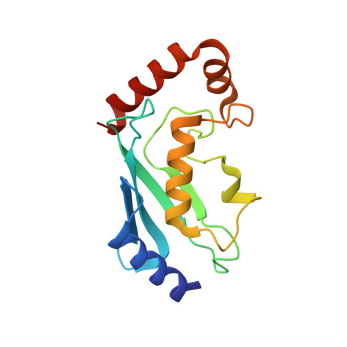
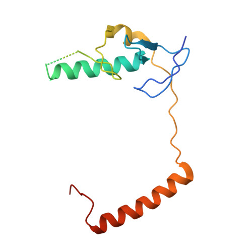
Insights into Ubiquitination from the Unique Clamp-like Binding of the RING E3 AO7 to the E2 UbcH5B.

Li, S.Liang, Y.H.Mariano, J.Metzger, M.B.Stringer, D.K.Hristova, V.A.Li, J.Randazzo, P.A.Tsai, Y.C.Ji, X.Weissman, A.M.

(2015) J Biological Chem 290: 30225-30239

  • DOI: https://doi.org/10.1074/jbc.M115.685867
  • Primary Citation Related Structures: 
    5D1K, 5D1L, 5D1M

  • PubMed Abstract: 

    RING proteins constitute the largest class of E3 ubiquitin ligases. Unlike most RINGs, AO7 (RNF25) binds the E2 ubiquitin-conjugating enzyme, UbcH5B (UBE2D2), with strikingly high affinity. We have defined, by co-crystallization, the distinctive means by which AO7 binds UbcH5B. AO7 contains a structurally unique UbcH5B binding region (U5BR) that is connected by an 11-amino acid linker to its RING domain, forming a clamp surrounding the E2. The U5BR interacts extensively with a region of UbcH5B that is distinct from both the active site and the RING-interacting region, referred to as the backside of the E2. An apparent paradox is that the high-affinity binding of the AO7 clamp to UbcH5B, which is dependent on the U5BR, decreases the rate of ubiquitination. We establish that this is a consequence of blocking the stimulatory, non-covalent, binding of ubiquitin to the backside of UbcH5B. Interestingly, when non-covalent backside ubiquitin binding cannot occur, the AO7 clamp now enhances the rate of ubiquitination. The high-affinity binding of the AO7 clamp to UbcH5B has also allowed for the co-crystallization of previously described and functionally important RING mutants at the RING-E2 interface. We show that mutations having marked effects on function only minimally affect the intermolecular interactions between the AO7 RING and UbcH5B, establishing a high degree of complexity in activation through the RING-E2 interface.


  • Organizational Affiliation
    • From the Laboratory of Protein Dynamics and Signaling.

Macromolecule Content 

  • Total Structure Weight: 32.86 kDa 
  • Atom Count: 2,494 
  • Modeled Residue Count: 256 
  • Deposited Residue Count: 280 
  • Unique protein chains: 2

Macromolecules

Find similar proteins by:|  3D Structure
Entity ID: 1
MoleculeChains  Sequence LengthOrganismDetailsImage
Ubiquitin-conjugating enzyme E2 D2147Homo sapiensMutation(s): 0 
Gene Names: UBE2D2PUBC1UBC4UBC5BUBCH4UBCH5B
EC: 6.3.2.19 (PDB Primary Data), 2.3.2.24 (UniProt), 2.3.2.23 (UniProt)
UniProt & NIH Common Fund Data Resources
Find proteins for P62837 (Homo sapiens)
Explore P62837 
Go to UniProtKB:  P62837
PHAROS:  P62837
GTEx:  ENSG00000131508 
Entity Groups
Sequence Clusters30% Identity50% Identity70% Identity90% Identity95% Identity100% Identity
UniProt GroupP62837
Sequence Annotations
Expand
Reference Sequence
Find similar proteins by:|  3D Structure
Entity ID: 2
MoleculeChains  Sequence LengthOrganismDetailsImage
E3 ubiquitin-protein ligase RNF25133Homo sapiensMutation(s): 0 
Gene Names: RNF25
EC: 6.3.2 (PDB Primary Data), 2.3.2.27 (UniProt)
UniProt & NIH Common Fund Data Resources
Find proteins for Q96BH1 (Homo sapiens)
Explore Q96BH1 
Go to UniProtKB:  Q96BH1
PHAROS:  Q96BH1
GTEx:  ENSG00000163481 
Entity Groups
Sequence Clusters30% Identity50% Identity70% Identity90% Identity95% Identity100% Identity
UniProt GroupQ96BH1
Sequence Annotations
Expand
Reference Sequence

Small Molecules

Ligands 4 Unique
IDChains Name / Formula / InChI Key2D Diagram3D Interactions
PEG

Query on PEG


Download:Ideal Coordinates CCD File
D [auth A]DI(HYDROXYETHYL)ETHER
C4 H10 O3
MTHSVFCYNBDYFN-UHFFFAOYSA-N
OXL

Query on OXL


Download:Ideal Coordinates CCD File
E [auth A]OXALATE ION
C2 O4
MUBZPKHOEPUJKR-UHFFFAOYSA-L
ZN

Query on ZN


Download:Ideal Coordinates CCD File
F [auth B],
G [auth B]
ZINC ION
Zn
PTFCDOFLOPIGGS-UHFFFAOYSA-N
EDO

Query on EDO


Download:Ideal Coordinates CCD File
C [auth A],
H [auth B],
I [auth B]
1,2-ETHANEDIOL
C2 H6 O2
LYCAIKOWRPUZTN-UHFFFAOYSA-N

Experimental Data & Validation

Experimental Data

  • Method: X-RAY DIFFRACTION
  • Resolution: 1.78 Å
  • R-Value Free:  0.202 (Depositor), 0.204 (DCC) 
  • R-Value Work:  0.170 (Depositor), 0.170 (DCC) 
  • R-Value Observed: 0.171 (Depositor) 
Space Group: C 1 2 1
Unit Cell:
Length ( Å )Angle ( ˚ )
a = 105.47α = 90
b = 45.658β = 118.09
c = 67.954γ = 90
Software Package:
Software NamePurpose
HKL-3000data reduction
PHENIXrefinement
PDB_EXTRACTdata extraction
HKL-3000data scaling
PHENIXphasing

Structure Validation

View Full Validation Report


Entry History 

Deposition Data

Revision History  (Full details and data files)

  • Version 1.0: 2015-10-28
    Type: Initial release
  • Version 1.1: 2015-12-30
    Changes: Database references
  • Version 1.2: 2023-08-30
    Changes: Data collection, Database references, Derived calculations, Structure summary
  • Version 1.3: 2023-09-27
    Changes: Refinement description