5D0M

Structure of UbE2D2:RNF165:Ub complex


Experimental Data Snapshot

  • Method: X-RAY DIFFRACTION
  • Resolution: 1.91 Å
  • R-Value Free: 0.213 
  • R-Value Work: 0.179 
  • R-Value Observed: 0.180 

wwPDB Validation   3D Report Full Report


This is version 1.2 of the entry. See complete history


Literature

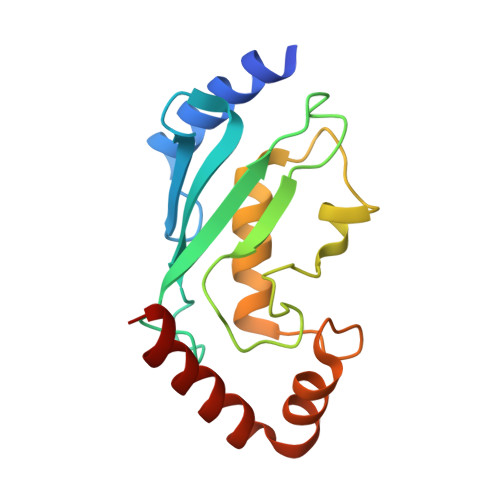
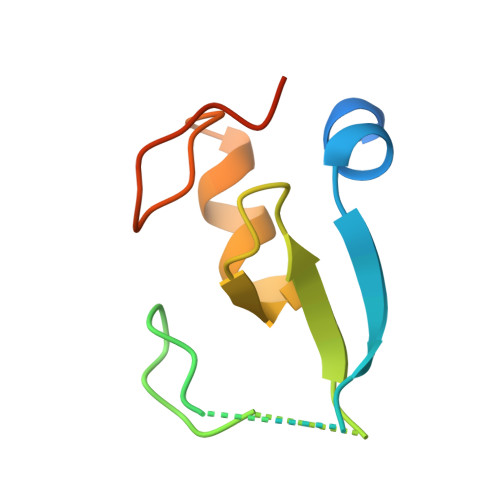
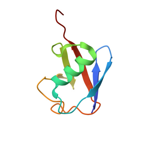
Secondary ubiquitin-RING docking enhances Arkadia and Ark2C E3 ligase activity.

Wright, J.D.Mace, P.D.Day, C.L.

(2016) Nat Struct Mol Biol 23: 45-52

  • DOI: https://doi.org/10.1038/nsmb.3142
  • Primary Citation of Related Structures:  
    5D0I, 5D0K, 5D0M

  • PubMed Abstract: 
  • RING-domain E3 ligases enhance transfer of ubiquitin to substrate proteins by stabilizing the RING-bound thioester-linked E2∼ubiquitin conjugate in a defined conformation that primes the active site for nucleophilic attack. Here we report that the monomeric RING domains from the human E3 ligases Arkadia and Ark2C bind directly to free ubiquitin with an affinity comparable to that of other dedicated ubiquitin-binding domains ...

    RING-domain E3 ligases enhance transfer of ubiquitin to substrate proteins by stabilizing the RING-bound thioester-linked E2∼ubiquitin conjugate in a defined conformation that primes the active site for nucleophilic attack. Here we report that the monomeric RING domains from the human E3 ligases Arkadia and Ark2C bind directly to free ubiquitin with an affinity comparable to that of other dedicated ubiquitin-binding domains. Further work showed that the Ark-like RING domain and the noncovalently bound ubiquitin molecule coordinately stabilize the E2-conjugated ubiquitin (donor ubiquitin) in the 'closed' conformation. Our studies identify the RING domain of Arkadia as a ubiquitin-binding domain and provide insight into a new ubiquitin-dependent mechanism used by monomeric RING domains to activate ubiquitin transfer. This study also suggests how substrates that have been monoubiquitinated could be favored for further ubiquitination.


    Organizational Affiliation

    Biochemistry Department, University of Otago, Dunedin, New Zealand.


Macromolecules
Find similar proteins by:  (by identity cutoff)  |  3D Structure
Entity ID: 1
MoleculeChainsSequence LengthOrganismDetailsImage
Ubiquitin-conjugating enzyme E2 D2152Homo sapiensMutation(s): 5 
Gene Names: UBE2D2PUBC1UBC4UBC5BUBCH4UBCH5B
EC: 6.3.2.19 (PDB Primary Data), 2.3.2.23 (UniProt), 2.3.2.24 (UniProt)
UniProt & NIH Common Fund Data Resources
Find proteins for P62837 (Homo sapiens)
Explore P62837 
Go to UniProtKB:  P62837
PHAROS:  P62837
Entity Groups  
Sequence Clusters30% Identity50% Identity70% Identity90% Identity95% Identity100% Identity
UniProt GroupP62837
Protein Feature View
Expand
  • Reference Sequence
Find similar proteins by:  (by identity cutoff)  |  3D Structure
Entity ID: 2
MoleculeChainsSequence LengthOrganismDetailsImage
RING finger protein 165B [auth C]87Homo sapiensMutation(s): 1 
Gene Names: RNF165
EC: 2.3.2.27
UniProt & NIH Common Fund Data Resources
Find proteins for Q6ZSG1 (Homo sapiens)
Explore Q6ZSG1 
Go to UniProtKB:  Q6ZSG1
PHAROS:  Q6ZSG1
Entity Groups  
Sequence Clusters30% Identity50% Identity70% Identity90% Identity95% Identity100% Identity
UniProt GroupQ6ZSG1
Protein Feature View
Expand
  • Reference Sequence
Find similar proteins by:  (by identity cutoff)  |  3D Structure
Entity ID: 3
MoleculeChainsSequence LengthOrganismDetailsImage
Polyubiquitin-BC [auth B]76Homo sapiensMutation(s): 0 
Gene Names: UBB
UniProt & NIH Common Fund Data Resources
Find proteins for P0CG47 (Homo sapiens)
Explore P0CG47 
Go to UniProtKB:  P0CG47
PHAROS:  P0CG47
Entity Groups  
Sequence Clusters30% Identity50% Identity70% Identity90% Identity95% Identity100% Identity
UniProt GroupP0CG47
Protein Feature View
Expand
  • Reference Sequence
Small Molecules
Ligands 2 Unique
IDChainsName / Formula / InChI Key2D Diagram3D Interactions
PO4
Query on PO4

Download Ideal Coordinates CCD File 
D [auth A]PHOSPHATE ION
O4 P
NBIIXXVUZAFLBC-UHFFFAOYSA-K
 Ligand Interaction
ZN
Query on ZN

Download Ideal Coordinates CCD File 
E [auth C],
F [auth C]
ZINC ION
Zn
PTFCDOFLOPIGGS-UHFFFAOYSA-N
 Ligand Interaction
Experimental Data & Validation

Experimental Data

  • Method: X-RAY DIFFRACTION
  • Resolution: 1.91 Å
  • R-Value Free: 0.213 
  • R-Value Work: 0.179 
  • R-Value Observed: 0.180 
  • Space Group: P 43
Unit Cell:
Length ( Å )Angle ( ˚ )
a = 62.62α = 90
b = 62.62β = 90
c = 82.25γ = 90
Software Package:
Software NamePurpose
PHENIXrefinement
iMOSFLMdata reduction
SCALAdata scaling
PHASERphasing

Structure Validation

View Full Validation Report


Entry History 

Deposition Data

Revision History  (Full details and data files)

  • Version 1.0: 2015-12-09
    Type: Initial release
  • Version 1.1: 2015-12-30
    Changes: Database references
  • Version 1.2: 2016-01-20
    Changes: Database references