5CYN | pdb_00005cyn

JC Virus large T-antigen origin binding domain F258L mutant


Experimental Data Snapshot

  • Method: X-RAY DIFFRACTION
  • Resolution: 2.70 Å
  • R-Value Free: 
    0.227 (Depositor), 0.241 (DCC) 
  • R-Value Work: 
    0.195 (Depositor), 0.205 (DCC) 
  • R-Value Observed: 
    0.197 (Depositor) 

Starting Model: experimental
View more details

wwPDB Validation 3D Report Full Report

Validation slider image for 5CYN

This is version 1.3 of the entry. See complete history

Literature

Structural Based Analyses of the JC Virus T-Antigen F258L Mutant Provides Evidence for DNA Dependent Conformational Changes in the C-Termini of Polyomavirus Origin Binding Domains.

Meinke, G.Phelan, P.J.Shin, J.Gagnon, D.Archambault, J.Bohm, A.Bullock, P.A.

(2016) PLoS Pathog 12: e1005362-e1005362

  • DOI: https://doi.org/10.1371/journal.ppat.1005362
  • Primary Citation Related Structures: 
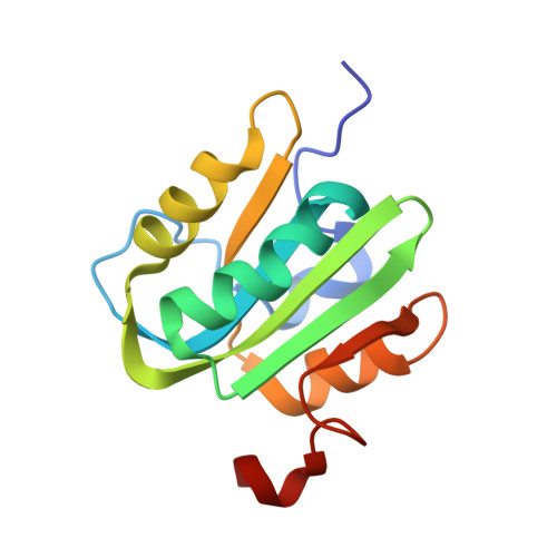
    5CYN

  • PubMed Abstract: 

    The replication of human polyomavirus JCV, which causes Progressive Multifocal Leukoencephalopathy, is initiated by the virally encoded T-antigen (T-ag). The structure of the JC virus T-ag origin-binding domain (OBD) was recently solved by X-ray crystallography. This structure revealed that the OBD contains a C-terminal pocket, and that residues from the multifunctional A1 and B2 motifs situated on a neighboring OBD molecule dock into the pocket. Related studies established that a mutation in a pocket residue (F258L) rendered JCV T-ag unable to support JCV DNA replication. To establish why this mutation inactivated JCV T-ag, we have solved the structure of the F258L JCV T-ag OBD mutant. Based on this structure, it is concluded that the structural consequences of the F258L mutation are limited to the pocket region. Further analyses, utilizing the available polyomavirus OBD structures, indicate that the F258 region is highly dynamic and that the relative positions of F258 are governed by DNA binding. The possible functional consequences of the DNA dependent rearrangements, including promotion of OBD cycling at the replication fork, are discussed.


  • Organizational Affiliation
    • Department of Developmental, Molecular and Chemical Biology, Tufts University School of Medicine, Boston, Massachusetts, United States of America.

Macromolecule Content 

  • Total Structure Weight: 15.05 kDa 
  • Atom Count: 1,043 
  • Modeled Residue Count: 127 
  • Deposited Residue Count: 130 
  • Unique protein chains: 1

Macromolecules

Find similar proteins by:|  3D Structure
Entity ID: 1
MoleculeChains  Sequence LengthOrganismDetailsImage
Large T antigen130JC polyomavirusMutation(s): 1 
EC: 3.6.4 (PDB Primary Data), 5.6.2.4 (UniProt)
UniProt
Find proteins for P03072 (JC polyomavirus)
Explore P03072 
Go to UniProtKB:  P03072
Entity Groups
Sequence Clusters30% Identity50% Identity70% Identity90% Identity95% Identity100% Identity
UniProt GroupP03072
Sequence Annotations
Expand
Reference Sequence

Small Molecules

Ligands 1 Unique
IDChains Name / Formula / InChI Key2D Diagram3D Interactions
EDO

Query on EDO


Download:Ideal Coordinates CCD File
B [auth A]1,2-ETHANEDIOL
C2 H6 O2
LYCAIKOWRPUZTN-UHFFFAOYSA-N

Experimental Data & Validation

Experimental Data

  • Method: X-RAY DIFFRACTION
  • Resolution: 2.70 Å
  • R-Value Free:  0.227 (Depositor), 0.241 (DCC) 
  • R-Value Work:  0.195 (Depositor), 0.205 (DCC) 
  • R-Value Observed: 0.197 (Depositor) 
Space Group: I 41
Unit Cell:
Length ( Å )Angle ( ˚ )
a = 103.54α = 90
b = 103.54β = 90
c = 34.34γ = 90
Software Package:
Software NamePurpose
REFMACrefinement
HKL-2000data reduction
SCALEPACKdata scaling
PHASERphasing

Structure Validation

View Full Validation Report


Entry History 

Deposition Data

Revision History  (Full details and data files)

  • Version 1.0: 2015-12-16
    Type: Initial release
  • Version 1.1: 2015-12-30
    Changes: Database references
  • Version 1.2: 2016-01-20
    Changes: Database references
  • Version 1.3: 2023-09-27
    Changes: Data collection, Database references, Derived calculations, Refinement description