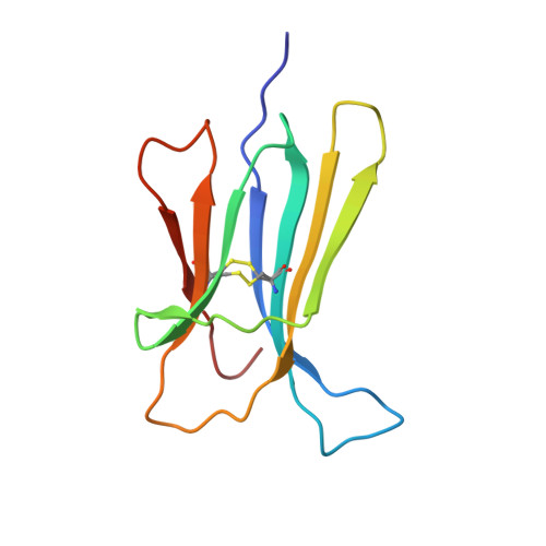
Conformational dynamics in crystals reveal the molecular bases for D76N beta-2 microglobulin aggregation propensity.
Le Marchand, T., de Rosa, M., Salvi, N., Sala, B.M., Andreas, L.B., Barbet-Massin, E., Sormanni, P., Barbiroli, A., Porcari, R., Sousa Mota, C., de Sanctis, D., Bolognesi, M., Emsley, L., Bellotti, V., Blackledge, M., Camilloni, C., Pintacuda, G., Ricagno, S.(2018) Nat Commun 9: 1658-1658
- PubMed: 29695721 Search on PubMedSearch on PubMed Central
- DOI: https://doi.org/10.1038/s41467-018-04078-y
- Primary Citation Related Structures:
4RMU, 4RMV, 4RMW, 5CS7, 5CSB, 5CSG - PubMed Abstract:
Spontaneous aggregation of folded and soluble native proteins in vivo is still a poorly understood process. A prototypic example is the D76N mutant of beta-2 microglobulin (β2m) that displays an aggressive aggregation propensity. Here we investigate the dynamics of β2m by X-ray crystallography, solid-state NMR, and molecular dynamics simulations to unveil the effects of the D76N mutation. Taken together, our data highlight the presence of minor disordered substates in crystalline β2m. The destabilization of the outer strands of D76N β2m accounts for the increased aggregation propensity. Furthermore, the computational modeling reveals a network of interactions with residue D76 as a keystone: this model allows predicting the stability of several point mutants. Overall, our study shows how the study of intrinsic dynamics in crystallo can provide crucial answers on protein stability and aggregation propensity. The comprehensive approach here presented may well be suited for the study of other folded amyloidogenic proteins.
- Centre de RMN à Très Hauts Champs, Institut des Sciences Analytiques (UMR 5280 CNRS/UCB Lyon 1/ENS Lyon), Université de Lyon, 69100, Villeurbanne, France.
Organizational Affiliation: