5CSF

S100B-RSK1 crystal structure A


Experimental Data Snapshot

  • Method: X-RAY DIFFRACTION
  • Resolution: 2.40 Å
  • R-Value Free: 0.291 
  • R-Value Work: 0.243 
  • R-Value Observed: 0.245 

wwPDB Validation   3D Report Full Report


This is version 1.2 of the entry. See complete history


Literature

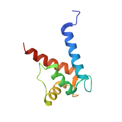
Structural Basis of Ribosomal S6 Kinase 1 (RSK1) Inhibition by S100B Protein: MODULATION OF THE EXTRACELLULAR SIGNAL-REGULATED KINASE (ERK) SIGNALING CASCADE IN A CALCIUM-DEPENDENT WAY.

Gogl, G.Alexa, A.Kiss, B.Katona, G.Kovacs, M.Bodor, A.Remenyi, A.Nyitray, L.

(2016) J Biol Chem 291: 11-27

  • DOI: https://doi.org/10.1074/jbc.M115.684928
  • Primary Citation of Related Structures:  
    5CSF, 5CSI, 5CSJ, 5CSN

  • PubMed Abstract: 
  • Mitogen-activated protein kinases (MAPK) promote MAPK-activated protein kinase activation. In the MAPK pathway responsible for cell growth, ERK2 initiates the first phosphorylation event on RSK1, which is inhibited by Ca(2+)-binding S100 proteins in malignant melanomas ...

    Mitogen-activated protein kinases (MAPK) promote MAPK-activated protein kinase activation. In the MAPK pathway responsible for cell growth, ERK2 initiates the first phosphorylation event on RSK1, which is inhibited by Ca(2+)-binding S100 proteins in malignant melanomas. Here, we present a detailed in vitro biochemical and structural characterization of the S100B-RSK1 interaction. The Ca(2+)-dependent binding of S100B to the calcium/calmodulin-dependent protein kinase (CaMK)-type domain of RSK1 is reminiscent of the better known binding of calmodulin to CaMKII. Although S100B-RSK1 and the calmodulin-CAMKII system are clearly distinct functionally, they demonstrate how unrelated intracellular Ca(2+)-binding proteins could influence the activity of the CaMK domain-containing protein kinases. Our crystallographic, small angle x-ray scattering, and NMR analysis revealed that S100B forms a "fuzzy" complex with RSK1 peptide ligands. Based on fast-kinetics experiments, we conclude that the binding involves both conformation selection and induced fit steps. Knowledge of the structural basis of this interaction could facilitate therapeutic targeting of melanomas.


    Organizational Affiliation

    From the Department of Biochemistry, nyitray@elte.hu.


Macromolecules
Find similar proteins by:  (by identity cutoff)  |  3D Structure
Entity ID: 1
MoleculeChainsSequence LengthOrganismDetailsImage
Protein S100-B
A, B
95Homo sapiensMutation(s): 0 
Gene Names: S100B
UniProt & NIH Common Fund Data Resources
Find proteins for P04271 (Homo sapiens)
Explore P04271 
Go to UniProtKB:  P04271
PHAROS:  P04271
Entity Groups  
Sequence Clusters30% Identity50% Identity70% Identity90% Identity95% Identity100% Identity
UniProt GroupP04271
Protein Feature View
Expand
  • Reference Sequence
Find similar proteins by:  (by identity cutoff)  |  3D Structure
Entity ID: 2
MoleculeChainsSequence LengthOrganismDetailsImage
Ribosomal protein S6 kinase alpha-155Homo sapiensMutation(s): 0 
Gene Names: RPS6KA1MAPKAPK1ARSK1
EC: 2.7.11.1
UniProt & NIH Common Fund Data Resources
Find proteins for Q15418 (Homo sapiens)
Explore Q15418 
Go to UniProtKB:  Q15418
PHAROS:  Q15418
Entity Groups  
Sequence Clusters30% Identity50% Identity70% Identity90% Identity95% Identity100% Identity
UniProt GroupQ15418
Protein Feature View
Expand
  • Reference Sequence
Small Molecules
Experimental Data & Validation

Experimental Data

  • Method: X-RAY DIFFRACTION
  • Resolution: 2.40 Å
  • R-Value Free: 0.291 
  • R-Value Work: 0.243 
  • R-Value Observed: 0.245 
  • Space Group: P 21 21 21
Unit Cell:
Length ( Å )Angle ( ˚ )
a = 38.3α = 90
b = 39.24β = 90
c = 172.82γ = 90
Software Package:
Software NamePurpose
PHENIXrefinement
XDSdata reduction
XDSdata scaling
PHASERphasing

Structure Validation

View Full Validation Report


Entry History 

Deposition Data

Revision History  (Full details and data files)

  • Version 1.0: 2015-11-11
    Type: Initial release
  • Version 1.1: 2015-11-18
    Changes: Database references
  • Version 1.2: 2016-01-13
    Changes: Database references