5CRW | pdb_00005crw

Crystal structure of the b'-a' domain of oxidized protein disulfide isomerase complexed with alpha-synuclein peptide (31-41)


Experimental Data Snapshot

  • Method: X-RAY DIFFRACTION
  • Resolution: 1.60 Å
  • R-Value Free: 
    0.217 (Depositor), 0.225 (DCC) 
  • R-Value Work: 
    0.184 (Depositor), 0.197 (DCC) 
  • R-Value Observed: 
    0.185 (Depositor) 

Starting Model: experimental
View more details

wwPDB Validation 3D Report Full Report

Validation slider image for 5CRW

This is version 1.4 of the entry. See complete history

Literature

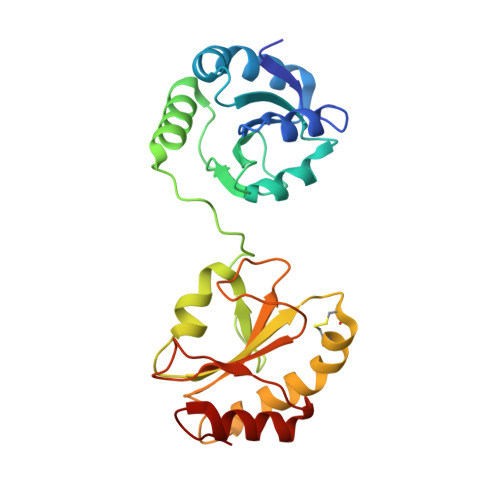
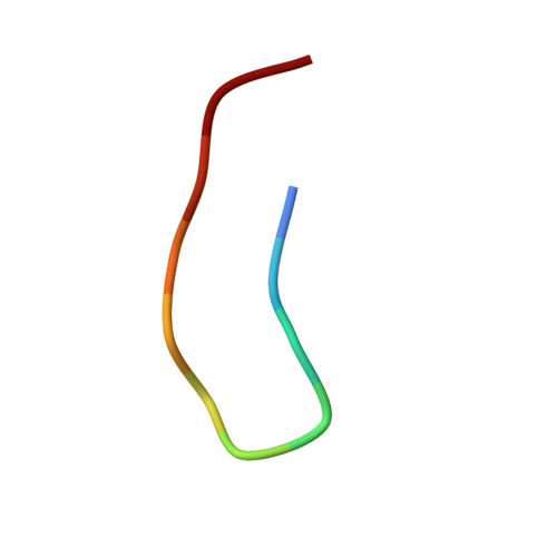
Structural basis of redox-dependent substrate binding of protein disulfide isomerase.

Yagi-Utsumi, M.Satoh, T.Kato, K.

(2015) Sci Rep 5: 13909-13909

  • DOI: https://doi.org/10.1038/srep13909
  • Primary Citation Related Structures: 
    5CRW

  • PubMed Abstract: 

    Protein disulfide isomerase (PDI) is a multidomain enzyme, operating as an essential folding catalyst, in which the b' and a' domains provide substrate binding sites and undergo an open-closed domain rearrangement depending on the redox states of the a' domain. Despite the long research history of this enzyme, three-dimensional structural data remain unavailable for its ligand-binding mode. Here we characterize PDI substrate recognition using α-synuclein (αSN) as the model ligand. Our nuclear magnetic resonance (NMR) data revealed that the substrate-binding domains of PDI captured the αSN segment Val37-Val40 only in the oxidized form. Furthermore, we determined the crystal structure of an oxidized form of the b'-a' domains in complex with an undecapeptide corresponding to this segment. The peptide-binding mode observed in the crystal structure with NMR validation, was characterized by hydrophobic interactions on the b' domain in an open conformation. Comparison with the previously reported crystal structure indicates that the a' domain partially masks the binding surface of the b' domain, causing steric hindrance against the peptide in the reduced form of the b'-a' domains that exhibits a closed conformation. These findings provide a structural basis for the mechanism underlying the redox-dependent substrate binding of PDI.


  • Organizational Affiliation
    • Okazaki Institute for Integrative Bioscience and Institute for Molecular Science, National Institutes of Natural Sciences, 5-1 Higashiyama, Myodaiji, Okazaki, Aichi 444-8787, Japan.

Macromolecule Content 

  • Total Structure Weight: 28.17 kDa 
  • Atom Count: 2,299 
  • Modeled Residue Count: 253 
  • Deposited Residue Count: 258 
  • Unique protein chains: 2

Macromolecules

Find similar proteins by:|  3D Structure
Entity ID: 1
MoleculeChains  Sequence LengthOrganismDetailsImage
Protein disulfide-isomerase247Mycothermus thermophilusMutation(s): 0 
EC: 5.3.4.1
UniProt
Find proteins for P55059 (Humicola insolens)
Explore P55059 
Go to UniProtKB:  P55059
Entity Groups
Sequence Clusters30% Identity50% Identity70% Identity90% Identity95% Identity100% Identity
UniProt GroupP55059
Sequence Annotations
Expand
Reference Sequence
Find similar proteins by:  Sequence   |   3D Structure  
Entity ID: 2
MoleculeChains  Sequence LengthOrganismDetailsImage
Peptide from Alpha-synuclein11Homo sapiensMutation(s): 0 
UniProt & NIH Common Fund Data Resources
Find proteins for P37840 (Homo sapiens)
Explore P37840 
Go to UniProtKB:  P37840
PHAROS:  P37840
GTEx:  ENSG00000145335 
Entity Groups
Sequence Clusters30% Identity50% Identity70% Identity90% Identity95% Identity100% Identity
UniProt GroupP37840
Sequence Annotations
Expand
Reference Sequence

Experimental Data & Validation

Experimental Data

  • Method: X-RAY DIFFRACTION
  • Resolution: 1.60 Å
  • R-Value Free:  0.217 (Depositor), 0.225 (DCC) 
  • R-Value Work:  0.184 (Depositor), 0.197 (DCC) 
  • R-Value Observed: 0.185 (Depositor) 
Space Group: P 21 21 21
Unit Cell:
Length ( Å )Angle ( ˚ )
a = 57.753α = 90
b = 62.51β = 90
c = 68.156γ = 90
Software Package:
Software NamePurpose
REFMACrefinement
DENZOdata reduction
SCALEPACKdata scaling
MOLREPphasing

Structure Validation

View Full Validation Report


Entry History 

& Funding Information

Deposition Data


Funding OrganizationLocationGrant Number
JSPS/MEXTJapan15K21680, 24770102, 25102001, 25102008, 25121730

Revision History  (Full details and data files)

  • Version 1.0: 2015-09-09
    Type: Initial release
  • Version 1.1: 2015-09-23
    Changes: Database references
  • Version 1.2: 2018-02-07
    Changes: Data collection, Database references, Derived calculations
  • Version 1.3: 2023-11-08
    Changes: Data collection, Database references, Refinement description
  • Version 1.4: 2024-10-16
    Changes: Structure summary