Direct observation of ultrafast collective motions in CO myoglobin upon ligand dissociation.
Barends, T.R., Foucar, L., Ardevol, A., Nass, K., Aquila, A., Botha, S., Doak, R.B., Falahati, K., Hartmann, E., Hilpert, M., Heinz, M., Hoffmann, M.C., Kofinger, J., Koglin, J.E., Kovacsova, G., Liang, M., Milathianaki, D., Lemke, H.T., Reinstein, J., Roome, C.M., Shoeman, R.L., Williams, G.J., Burghardt, I., Hummer, G., Boutet, S., Schlichting, I.(2015) Science 350: 445-450
- PubMed: 26359336 Search on PubMed
- DOI: https://doi.org/10.1126/science.aac5492
- Primary Citation Related Structures:
5CMV, 5CN4, 5CN5, 5CN6, 5CN7, 5CN8, 5CN9, 5CNB, 5CNC, 5CND, 5CNE, 5CNF, 5CNG, 5D5R - PubMed Abstract:
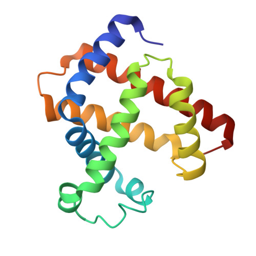
The hemoprotein myoglobin is a model system for the study of protein dynamics. We used time-resolved serial femtosecond crystallography at an x-ray free-electron laser to resolve the ultrafast structural changes in the carbonmonoxy myoglobin complex upon photolysis of the Fe-CO bond. Structural changes appear throughout the protein within 500 femtoseconds, with the C, F, and H helices moving away from the heme cofactor and the E and A helices moving toward it. These collective movements are predicted by hybrid quantum mechanics/molecular mechanics simulations. Together with the observed oscillations of residues contacting the heme, our calculations support the prediction that an immediate collective response of the protein occurs upon ligand dissociation, as a result of heme vibrational modes coupling to global modes of the protein.
- Max-Planck-Institut für Medizinische Forschung, Jahnstraße 29, 69120 Heidelberg, Germany. thomas.barends@mpimf-heidelberg.mpg.de ilme.schlichting@mpimf-heidelberg.mpg.de.
Organizational Affiliation: