5CJ0

Crystal Structure of Amino Acids 1631-1692 of MYH7


Experimental Data Snapshot

  • Method: X-RAY DIFFRACTION
  • Resolution: 2.30 Å
  • R-Value Free: 0.238 
  • R-Value Work: 0.216 
  • R-Value Observed: 0.217 

wwPDB Validation   3D Report Full Report


This is version 1.4 of the entry. See complete history


Literature

A composite approach towards a complete model of the myosin rod.

Korkmaz, E.N.Taylor, K.C.Andreas, M.P.Ajay, G.Heinze, N.T.Cui, Q.Rayment, I.

(2016) Proteins 84: 172-189

  • DOI: https://doi.org/10.1002/prot.24964
  • Primary Citation of Related Structures:  
    5CHX, 5CJ0, 5CJ1, 5CJ4

  • PubMed Abstract: 

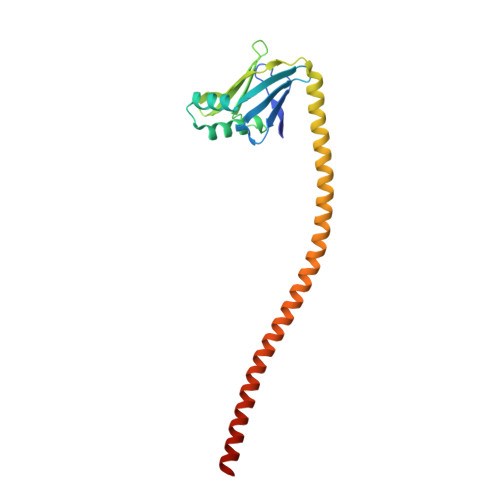
    Sarcomeric myosins have the remarkable ability to form regular bipolar thick filaments that, together with actin thin filaments, constitute the fundamental contractile unit of skeletal and cardiac muscle. This has been established for over 50 years and yet a molecular model for the thick filament has not been attained. In part this is due to the lack of a detailed molecular model for the coiled-coil that constitutes the myosin rod. The ability to self-assemble resides in the C-terminal section of myosin known as light meromyosin (LMM) which exhibits strong salt-dependent aggregation that has inhibited structural studies. Here we evaluate the feasibility of generating a complete model for the myosin rod by combining overlapping structures of five sections of coiled-coil covering 164 amino acid residues which constitute 20% of LMM. Each section contains ∼ 7-9 heptads of myosin. The problem of aggregation was overcome by incorporating the globular folding domains, Gp7 and Xrcc4 which enhance crystallization. The effect of these domains on the stability and conformation of the myosin rod was examined through biophysical studies and overlapping structures. In addition, a computational approach was developed to combine the sections into a contiguous model. The structures were aligned, trimmed to form a contiguous model, and simulated for >700 ns to remove the discontinuities and achieve an equilibrated conformation that represents the native state. This experimental and computational strategy lays the foundation for building a model for the entire myosin rod.


  • Organizational Affiliation

    Department of Chemistry and Theoretical Chemistry Institute, University of Wisconsin, Madison, WI 53706, USA.


Macromolecules
Find similar proteins by:  (by identity cutoff)  |  3D Structure
Entity ID: 1
MoleculeChains Sequence LengthOrganismDetailsImage
Xrcc4-MYH7-(1631-1692) chimera protein
A, B
207Homo sapiensMutation(s): 1 
Gene Names: XRCC4MYH7MYHCB
UniProt & NIH Common Fund Data Resources
Find proteins for Q13426 (Homo sapiens)
Explore Q13426 
Go to UniProtKB:  Q13426
PHAROS:  Q13426
GTEx:  ENSG00000152422 
Find proteins for P12883 (Homo sapiens)
Explore P12883 
Go to UniProtKB:  P12883
PHAROS:  P12883
GTEx:  ENSG00000092054 
Entity Groups  
Sequence Clusters30% Identity50% Identity70% Identity90% Identity95% Identity100% Identity
UniProt GroupsP12883Q13426
Sequence Annotations
Expand
  • Reference Sequence
Experimental Data & Validation

Experimental Data

  • Method: X-RAY DIFFRACTION
  • Resolution: 2.30 Å
  • R-Value Free: 0.238 
  • R-Value Work: 0.216 
  • R-Value Observed: 0.217 
  • Space Group: C 1 2 1
Unit Cell:
Length ( Å )Angle ( ˚ )
a = 200.006α = 90
b = 44.355β = 108.19
c = 74.664γ = 90
Software Package:
Software NamePurpose
PHENIXrefinement
PDB_EXTRACTdata extraction
HKL-3000data reduction
HKL-3000data scaling
PHENIXphasing

Structure Validation

View Full Validation Report


Entry History & Funding Information

Deposition Data


Funding OrganizationLocationGrant Number
National Institutes of Health/National Heart, Lung, and Blood Institute (NIH/NHLBI)United StatesHL111237
National Science Foundation (NSF, United States)United StatesCHE1300209

Revision History  (Full details and data files)

  • Version 1.0: 2015-12-02
    Type: Initial release
  • Version 1.1: 2016-01-27
    Changes: Database references
  • Version 1.2: 2017-09-27
    Changes: Author supporting evidence, Database references, Derived calculations
  • Version 1.3: 2019-11-27
    Changes: Author supporting evidence
  • Version 1.4: 2023-09-27
    Changes: Data collection, Database references, Refinement description