5CHA

THE REFINEMENT AND THE STRUCTURE OF THE DIMER OF ALPHA-*CHYMOTRYPSIN AT 1.67-*ANGSTROMS RESOLUTION


Experimental Data Snapshot

  • Method: X-RAY DIFFRACTION
  • Resolution: 1.67 Å

wwPDB Validation   3D Report Full Report


This is version 1.2 of the entry. See complete history


Literature

The refinement and the structure of the dimer of alpha-chymotrypsin at 1.67-A resolution.

Blevins, R.A.Tulinsky, A.

(1985) J Biol Chem 260: 4264-4275

  • DOI: https://doi.org/10.2210/pdb5cha/pdb
  • Primary Citation of Related Structures:  
    5CHA

  • PubMed Abstract: 

    The two molecules of the asymmetric unit of the pH 3.5 conformer of alpha-chymotrypsin have been refined at 1.67-A resolution using restrained least squares methods with Hendrickson's program (PROLSQ). The final R factor is 0.179 (including 247 water molecules). The folding of the main chain of the independent molecules is the same within experimental error but the same does not generally apply to the side chain stereochemistry. From this we conclude that the folding of a protein structure is basically independent of most of the detailed stereochemistry of its side chains. The side chains of the interface region between the independent molecules display pronounced asymmetry. This asymmetry suggests that dynamic and asymmetrical structural changes take place at the time of oligomerization leading to more energetically favorable interactions for the dimer. Comparison of the structures of the independent molecules of alpha-chymotrypsin with the structure of monomeric gamma-chymotrypsin revealed that although the folding of the three molecules is essentially the same, numerous and significant differences pervade the side chain stereochemistry attributable to general flexibility. The specificity site of alpha-chymotrypsin is occupied by ordered water molecules in a similar way to gamma-chymotrypsin and other proteins. Some of these water molecules are displaced when substrate binds to the enzyme, while the others appear to help identify and position the aromatic side chain in catalysis.


Macromolecules

Find similar proteins by:  Sequence   |   3D Structure  

Entity ID: 1
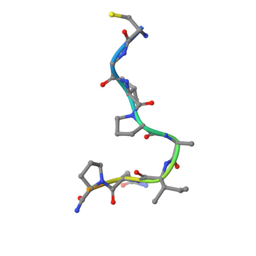
MoleculeChains Sequence LengthOrganismDetailsImage
ALPHA-CHYMOTRYPSIN AA,
D [auth E]
13Bos taurusMutation(s): 0 
EC: 3.4.21.1
UniProt
Find proteins for P00766 (Bos taurus)
Explore P00766 
Go to UniProtKB:  P00766
Entity Groups  
Sequence Clusters30% Identity50% Identity70% Identity90% Identity95% Identity100% Identity
UniProt GroupP00766
Sequence Annotations
Expand
  • Reference Sequence
Find similar proteins by:  (by identity cutoff)  |  3D Structure
Entity ID: 2
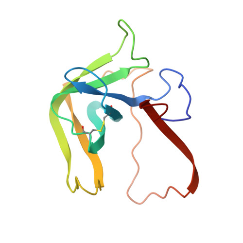
MoleculeChains Sequence LengthOrganismDetailsImage
ALPHA-CHYMOTRYPSIN AB,
E [auth F]
131Bos taurusMutation(s): 0 
EC: 3.4.21.1
UniProt
Find proteins for P00766 (Bos taurus)
Explore P00766 
Go to UniProtKB:  P00766
Entity Groups  
Sequence Clusters30% Identity50% Identity70% Identity90% Identity95% Identity100% Identity
UniProt GroupP00766
Sequence Annotations
Expand
  • Reference Sequence
Find similar proteins by:  (by identity cutoff)  |  3D Structure
Entity ID: 3
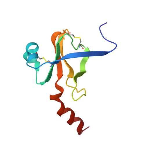
MoleculeChains Sequence LengthOrganismDetailsImage
ALPHA-CHYMOTRYPSIN AC,
F [auth G]
97Bos taurusMutation(s): 0 
EC: 3.4.21.1
UniProt
Find proteins for P00766 (Bos taurus)
Explore P00766 
Go to UniProtKB:  P00766
Entity Groups  
Sequence Clusters30% Identity50% Identity70% Identity90% Identity95% Identity100% Identity
UniProt GroupP00766
Sequence Annotations
Expand
  • Reference Sequence
Experimental Data & Validation

Experimental Data

  • Method: X-RAY DIFFRACTION
  • Resolution: 1.67 Å
  • Space Group: P 1 21 1
Unit Cell:
Length ( Å )Angle ( ˚ )
a = 49.29α = 90
b = 67.48β = 102.02
c = 65.94γ = 90

Structure Validation

View Full Validation Report


Entry History 

Deposition Data

Revision History  (Full details and data files)

  • Version 1.0: 1985-04-01
    Type: Initial release
  • Version 1.1: 2008-03-03
    Changes: Version format compliance
  • Version 1.2: 2011-07-13
    Changes: Version format compliance