5CGX | pdb_00005cgx

CRYSTAL STRUCTURE OF Fox-4 cephamycinase mutant Y150F complexed with cefoxitin


Experimental Data Snapshot

  • Method: X-RAY DIFFRACTION
  • Resolution: 1.21 Å
  • R-Value Free: 
    0.205 (Depositor), 0.205 (DCC) 
  • R-Value Work: 
    0.181 (Depositor), 0.183 (DCC) 
  • R-Value Observed: 
    0.182 (Depositor) 

Starting Model: experimental
View more details

wwPDB Validation 3D Report Full Report

Validation slider image for 5CGX

Ligand Structure Quality Assessment 


This is version 1.2 of the entry. See complete history

Literature

FOX-4 cephamycinase: an analysis of structure and function.

Lefurgy, S.T.Malashkevich, V.N.Aguilan, J.T.Nieves, E.Mundorff, E.C.Biju, B.Noel, M.A.Toro, R.Baiwir, D.Papp-Wallace, K.M.Almo, S.C.Frere, J.M.Bou, G.Bonomo, R.A.

(2015) Antimicrob Agents Chemother 

  • DOI: https://doi.org/10.1128/AAC.01887-15
  • Primary Citation Related Structures: 
    5CGS, 5CGW, 5CGX

  • PubMed Abstract: 

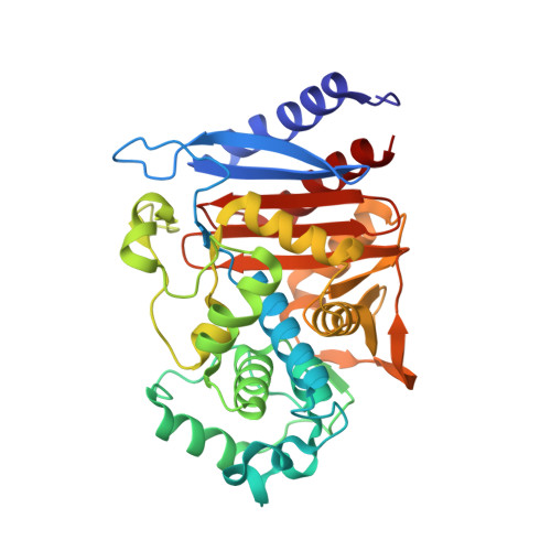
    Class C β-lactamases poorly hydrolyze cephamycins (e.g., cefoxitin, cefotetan, and moxalactam). In the past 2 decades, a new family of plasmid-based AmpC β-lactamases conferring resistance to cefoxitin, the FOX family, has grown to include nine unique members descended from the Aeromonas caviae chromosomal AmpC. To understand the basis for the unique cephamycinase activity in the FOX family, we determined the first X-ray crystal structures of FOX-4, apo enzyme and the acyl-enzyme with its namesake compound, cefoxitin, using the Y150F deacylation-deficient variant. Notably, recombinant expression of N-terminally tagged FOX-4 also yielded an inactive adenylylated enzyme form not previously observed in β-lactamases. The posttranslational modification (PTM), which occurs on the active site Ser64, would not seem to provide a selective advantage, yet might present an opportunity for the design of novel antibacterial drugs. Substantial ligand-induced changes in the enzyme are seen in the acyl-enzyme complex, particularly the R2 loop and helix H10 (P289 to N297), with movement of F293 by 10.3 Å. Taken together, this study provides the first picture of this highly proficient class C cephamycinase, uncovers a novel PTM, and suggests a possible cephamycin resistance mechanism involving repositioning of the substrate due to the presence of S153P, N289P, and N346I substitutions in the ligand binding pocket.


  • Organizational Affiliation
    • Department of Chemistry, Hofstra University, Hempstead, New York, USA.

Macromolecule Content 

  • Total Structure Weight: 39.58 kDa 
  • Atom Count: 3,175 
  • Modeled Residue Count: 362 
  • Deposited Residue Count: 362 
  • Unique protein chains: 1

Macromolecules

Find similar proteins by:|  3D Structure
Entity ID: 1
MoleculeChains  Sequence LengthOrganismDetailsImage
Beta-lactamase362Escherichia coliMutation(s): 1 
Gene Names: fox-4
EC: 3.5.2.6
UniProt
Find proteins for Q9L387 (Escherichia coli)
Explore Q9L387 
Go to UniProtKB:  Q9L387
Entity Groups
Sequence Clusters30% Identity50% Identity70% Identity90% Identity95% Identity100% Identity
UniProt GroupQ9L387
Sequence Annotations
Expand
Reference Sequence

Small Molecules

Ligands 3 Unique
IDChains Name / Formula / InChI Key2D Diagram3D Interactions
1S7

Query on 1S7


Download:Ideal Coordinates CCD File
B [auth A](2R)-2-{(1S)-1-methoxy-2-oxo-1-[(thiophen-2-ylacetyl)amino]ethyl}-5-methylidene-5,6-dihydro-2H-1,3-thiazine-4-carboxylic acid
C15 H16 N2 O5 S2
BMQDZZSQSZJJQV-CABCVRRESA-N
ZN

Query on ZN


Download:Ideal Coordinates CCD File
C [auth A]
D [auth A]
E [auth A]
F [auth A]
G [auth A]
C [auth A],
D [auth A],
E [auth A],
F [auth A],
G [auth A],
I [auth A]
ZINC ION
Zn
PTFCDOFLOPIGGS-UHFFFAOYSA-N
NA

Query on NA


Download:Ideal Coordinates CCD File
H [auth A]SODIUM ION
Na
FKNQFGJONOIPTF-UHFFFAOYSA-N

Experimental Data & Validation

Experimental Data

  • Method: X-RAY DIFFRACTION
  • Resolution: 1.21 Å
  • R-Value Free:  0.205 (Depositor), 0.205 (DCC) 
  • R-Value Work:  0.181 (Depositor), 0.183 (DCC) 
  • R-Value Observed: 0.182 (Depositor) 
Space Group: P 1 21 1
Unit Cell:
Length ( Å )Angle ( ˚ )
a = 54.524α = 90
b = 56.588β = 99.23
c = 55.485γ = 90
Software Package:
Software NamePurpose
PHASERphasing
PHENIXrefinement
PDB_EXTRACTdata extraction
MOSFLMdata reduction
SCALAdata scaling
PHASERphasing

Structure Validation

View Full Validation Report


Ligand Structure Quality Assessment 


Entry History 

Deposition Data

Revision History  (Full details and data files)

  • Version 1.0: 2016-02-03
    Type: Initial release
  • Version 1.1: 2023-09-27
    Changes: Data collection, Database references, Derived calculations, Refinement description
  • Version 1.2: 2024-10-23
    Changes: Structure summary