5CFZ | pdb_00005cfz

Crystal structure of E. coli FabI in apo form


Experimental Data Snapshot

  • Method: X-RAY DIFFRACTION
  • Resolution: 1.97 Å
  • R-Value Free: 
    0.225 (Depositor), 0.222 (DCC) 
  • R-Value Work: 
    0.190 (Depositor), 0.188 (DCC) 
  • R-Value Observed: 
    0.192 (Depositor) 

Starting Model: experimental
View more details

wwPDB Validation 3D Report Full Report

Validation slider image for 5CFZ

This is version 1.1 of the entry. See complete history

Literature

Crystallographic insights into the structure-activity relationships of diazaborine enoyl-ACP reductase inhibitors.

Jordan, C.A.Sandoval, B.A.Serobyan, M.V.Gilling, D.H.Groziak, M.P.Xu, H.H.Vey, J.L.

(2015) Acta Crystallogr F Struct Biol Commun 71: 1521-1530

  • DOI: https://doi.org/10.1107/S2053230X15022098
  • Primary Citation Related Structures: 
    5CFZ, 5CG1, 5CG2

  • PubMed Abstract: 

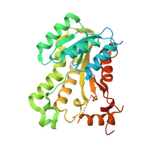
    Enoyl-ACP reductase, the last enzyme of the fatty-acid biosynthetic pathway, is the molecular target for several successful antibiotics such as the tuberculosis therapeutic isoniazid. It is currently under investigation as a narrow-spectrum antibiotic target for the treatment of several types of bacterial infections. The diazaborine family is a group of boron heterocycle-based synthetic antibacterial inhibitors known to target enoyl-ACP reductase. Development of this class of molecules has thus far focused solely on the sulfonyl-containing versions. Here, the requirement for the sulfonyl group in the diazaborine scaffold was investigated by examining several recently characterized enoyl-ACP reductase inhibitors that lack the sulfonyl group and exhibit additional variability in substitutions, size and flexibility. Biochemical studies are reported showing the inhibition of Escherichia coli enoyl-ACP reductase by four diazaborines, and the crystal structures of two of the inhibitors bound to E. coli enoyl-ACP reductase solved to 2.07 and 2.11 Å resolution are reported. The results show that the sulfonyl group can be replaced with an amide or thioamide without disruption of the mode of inhibition of the molecule.


  • Organizational Affiliation
    • Department of Chemistry and Biochemistry, California State University Northridge, Northridge, CA 91330-8262, USA.

Macromolecule Content 

  • Total Structure Weight: 65.38 kDa 
  • Atom Count: 3,879 
  • Modeled Residue Count: 495 
  • Deposited Residue Count: 610 
  • Unique protein chains: 1

Macromolecules

Find similar proteins by:|  3D Structure
Entity ID: 1
MoleculeChains  Sequence LengthOrganismDetailsImage
Enoyl-[acyl-carrier-protein] reductase [NADH] FabI
A, B
305Escherichia coli K-12Mutation(s): 0 
Gene Names: fabIenvMb1288JW1281
EC: 1.3.1.9
UniProt
Find proteins for P0AEK4 (Escherichia coli (strain K12))
Explore P0AEK4 
Go to UniProtKB:  P0AEK4
Entity Groups
Sequence Clusters30% Identity50% Identity70% Identity90% Identity95% Identity100% Identity
UniProt GroupP0AEK4
Sequence Annotations
Expand
Reference Sequence

Experimental Data & Validation

Experimental Data

  • Method: X-RAY DIFFRACTION
  • Resolution: 1.97 Å
  • R-Value Free:  0.225 (Depositor), 0.222 (DCC) 
  • R-Value Work:  0.190 (Depositor), 0.188 (DCC) 
  • R-Value Observed: 0.192 (Depositor) 
Space Group: P 61 2 2
Unit Cell:
Length ( Å )Angle ( ˚ )
a = 80.152α = 90
b = 80.152β = 90
c = 323.94γ = 120
Software Package:
Software NamePurpose
REFMACrefinement
SCALAdata scaling
PDB_EXTRACTdata extraction
XDSdata reduction
PHASERphasing

Structure Validation

View Full Validation Report


Entry History 

& Funding Information

Deposition Data


Funding OrganizationLocationGrant Number
CSUN Faculty DevelopmentUnited StatesProbationary Faculty Support Grant

Revision History  (Full details and data files)

  • Version 1.0: 2015-12-09
    Type: Initial release
  • Version 1.1: 2023-09-27
    Changes: Data collection, Database references, Derived calculations, Refinement description