5CCB

Crystal structure of human m1A58 methyltransferase in a complex with tRNA3Lys and SAH


Experimental Data Snapshot

  • Method: X-RAY DIFFRACTION
  • Resolution: 2.00 Å
  • R-Value Free: 0.222 
  • R-Value Work: 0.199 
  • R-Value Observed: 0.200 

wwPDB Validation   3D Report Full Report


Ligand Structure Quality Assessment 


This is version 1.4 of the entry. See complete history


Literature

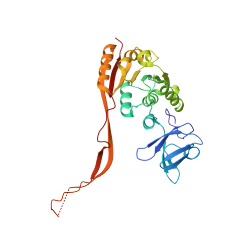
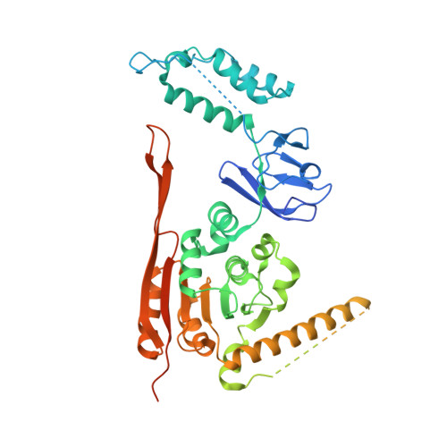
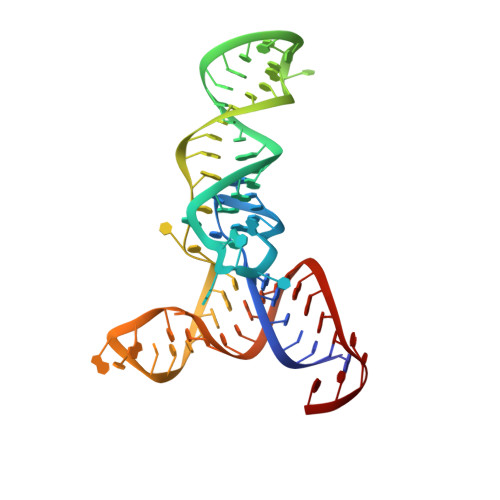
Crystal Structure of the Human tRNA m(1)A58 Methyltransferase-tRNA3(Lys) Complex: Refolding of Substrate tRNA Allows Access to the Methylation Target.

Finer-Moore, J.Czudnochowski, N.O'Connell, J.D.Wang, A.L.Stroud, R.M.

(2015) J Mol Biol 427: 3862-3876

  • DOI: https://doi.org/10.1016/j.jmb.2015.10.005
  • Primary Citation of Related Structures:  
    5CCB, 5CCX, 5CD1

  • PubMed Abstract: 

    Human tRNA3(Lys) is the primer for reverse transcription of HIV; the 3' end is complementary to the primer-binding site on HIV RNA. The complementarity ends at the 18th base, A58, which in tRNA3(Lys) is modified to remove Watson-Crick pairing. Motivated to test the role of the modification in terminating the primer-binding sequence and thus limiting run-on transcription, we asked how the modification of RNA could be accomplished. tRNA m(1)A58 methyltransferase (m(1)A58 MTase) methylates N1 of A58, which is buried in the TΨC-loop of tRNA, from cofactor S-adenosyl-L-methionine. This conserved tRNA modification is essential for stability of initiator tRNA in Saccharomyces cerevisiae. Reported here, three structures of human tRNA m(1)A58 MTase in complex with human tRNA3(Lys) and the product S-adenosyl-L-homocysteine show a dimer of heterodimers in which each heterodimer comprises a catalytic chain, Trm61, and a homologous but noncatalytic chain, Trm6, repurposed as a tRNA-binding subunit that acts in trans; tRNAs bind across the dimer interface such that Trm6 from the opposing heterodimer brings A58 into the active site of Trm61. T-loop and D-loop are splayed apart showing how A58, normally buried in tRNA, becomes accessible for modification. This result has broad impact on our understanding of the mechanisms of modifying internal sites in folded tRNA. The structures serve as templates for design of inhibitors that could be used to test tRNA m(1)A58 MTase's impact on retroviral priming and transcription.


  • Organizational Affiliation

    Department of Biochemistry and Biophysics, University of California San Francisco, San Francisco, CA 94143, USA. Electronic address: finer@msg.ucsf.edu.


Macromolecules

Find similar proteins by:  (by identity cutoff)  |  3D Structure
Entity ID: 1
MoleculeChains Sequence LengthOrganismDetailsImage
tRNA (adenine(58)-N(1))-methyltransferase catalytic subunit TRMT61A289Homo sapiensMutation(s): 0 
Gene Names: TRMT61AC14orf172TRM61
EC: 2.1.1.220
UniProt & NIH Common Fund Data Resources
Find proteins for Q96FX7 (Homo sapiens)
Explore Q96FX7 
Go to UniProtKB:  Q96FX7
PHAROS:  Q96FX7
GTEx:  ENSG00000166166 
Entity Groups  
Sequence Clusters30% Identity50% Identity70% Identity90% Identity95% Identity100% Identity
UniProt GroupQ96FX7
Sequence Annotations
Expand
  • Reference Sequence
Find similar proteins by:  (by identity cutoff)  |  3D Structure
Entity ID: 2
MoleculeChains Sequence LengthOrganismDetailsImage
tRNA (adenine(58)-N(1))-methyltransferase non-catalytic subunit TRM6497Homo sapiensMutation(s): 0 
Gene Names: TRMT6KIAA1153TRM6CGI-09
EC: 2.1.1.220
UniProt & NIH Common Fund Data Resources
Find proteins for Q9UJA5 (Homo sapiens)
Explore Q9UJA5 
Go to UniProtKB:  Q9UJA5
PHAROS:  Q9UJA5
GTEx:  ENSG00000089195 
Entity Groups  
Sequence Clusters30% Identity50% Identity70% Identity90% Identity95% Identity100% Identity
UniProt GroupQ9UJA5
Sequence Annotations
Expand
  • Reference Sequence
Find similar nucleic acids by:  (by identity cutoff)  |  3D Structure
Entity ID: 3
MoleculeChains LengthOrganismImage
tRNA3LysC [auth N]77Homo sapiens
Sequence Annotations
Expand
  • Reference Sequence
Small Molecules
Ligands 2 Unique
IDChains Name / Formula / InChI Key2D Diagram3D Interactions
SAH
Query on SAH

Download Ideal Coordinates CCD File 
D [auth A]S-ADENOSYL-L-HOMOCYSTEINE
C14 H20 N6 O5 S
ZJUKTBDSGOFHSH-WFMPWKQPSA-N
NA
Query on NA

Download Ideal Coordinates CCD File 
E [auth N]SODIUM ION
Na
FKNQFGJONOIPTF-UHFFFAOYSA-N
Experimental Data & Validation

Experimental Data

  • Method: X-RAY DIFFRACTION
  • Resolution: 2.00 Å
  • R-Value Free: 0.222 
  • R-Value Work: 0.199 
  • R-Value Observed: 0.200 
  • Space Group: P 43 21 2
Unit Cell:
Length ( Å )Angle ( ˚ )
a = 137.18α = 90
b = 137.18β = 90
c = 177.02γ = 90
Software Package:
Software NamePurpose
PHENIXrefinement
XDSdata reduction
XSCALEdata scaling

Structure Validation

View Full Validation Report


Ligand Structure Quality Assessment 


Entry History & Funding Information

Deposition Data


Funding OrganizationLocationGrant Number
National Institutes of Health/National Institute of General Medical Sciences (NIH/NIGMS)United StatesGM51232
National Institutes of Health/National Institute of General Medical Sciences (NIH/NIGMS)United StatesGM0082250
German Research Foundation (DFG)GermanyCZ 205/1-1

Revision History  (Full details and data files)

  • Version 1.0: 2015-11-04
    Type: Initial release
  • Version 1.1: 2015-12-16
    Changes: Database references
  • Version 1.2: 2017-09-27
    Changes: Author supporting evidence, Database references, Derived calculations
  • Version 1.3: 2019-12-25
    Changes: Author supporting evidence, Database references
  • Version 1.4: 2023-09-27
    Changes: Data collection, Database references, Derived calculations, Refinement description