5CB5

Structural Insights into the Mechanism of Escherichia coli Ymdb


Experimental Data Snapshot

  • Method: X-RAY DIFFRACTION
  • Resolution: 2.80 Å
  • R-Value Free: 0.290 
  • R-Value Work: 0.262 
  • R-Value Observed: 0.263 

wwPDB Validation   3D Report Full Report


This is version 1.2 of the entry. See complete history


Literature

Structural insights into the mechanism of Escherichia coli YmdB: A 2'-O-acetyl-ADP-ribose deacetylase

Zhang, W.Wang, C.Song, Y.Shao, C.Zhang, X.Zang, J.

(2015) J Struct Biol 192: 478-486

  • DOI: https://doi.org/10.1016/j.jsb.2015.10.010
  • Primary Citation of Related Structures:  
    5CB3, 5CB5, 5CMS

  • PubMed Abstract: 

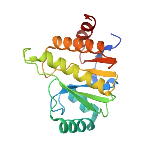
    The Escherichia coli protein YmdB belongs to the macrodomain protein family, which can bind ADP-ribose (ADPr) and its derivatives. Recently, YmdB was reported to be capable of deacetylating O-acetyl-ADP-ribose (OAADPr) to yield ADPr and free acetate. To study the substrate specificity and catalytic mechanism, the crystal structures of E. coli YmdB in complex with ADPr, double mutant N25AD35A complexed with 2'-OAADPr, and Y126A/ADPr complex were solved at 1.8Å, 2.8Å and 3.0Å resolution, respectively. Structural and biochemical studies reveal that YmdB has substrate specificity against 2'-OAADPr. The conserved residues Asn25 and Asp35 are crucial for catalytic activity, and an active water molecule is proposed as the nucleophile to attack the acetyl group of 2'-OAADPr. Our findings indicate that the conserved phenyl group of Tyr126 plays a crucial role in catalytic activity by stabilizing the right orientation of distal ribose and that Gly32 may be important for activity by interacting with the acetyl group of 2'-OAADPr. Based on these observations, a model of YmdB in complex with 2'-OAADPr was made to illustrate the proposed catalytic mechanism of YmdB.


  • Organizational Affiliation

    Hefei National Laboratory for Physical Sciences at Microscale, School of Life Sciences, University of Science and Technology of China, 96 Jinzhai Road, Hefei, Anhui 230026, People's Republic of China; Key Laboratory of Structural Biology, Chinese Academy of Sciences, Hefei, Anhui 230027, People's Republic of China; National Synchrotron Radiation Laboratory, University of Science and Technology of China, Hefei, Anhui 230027, People's Republic of China.


Macromolecules
Find similar proteins by:  (by identity cutoff)  |  3D Structure
Entity ID: 1
MoleculeChains Sequence LengthOrganismDetailsImage
O-acetyl-ADP-ribose deacetylase183Escherichia coli K-12Mutation(s): 2 
Gene Names: ymdB
EC: 3.5.1
UniProt
Find proteins for P0A8D6 (Escherichia coli (strain K12))
Explore P0A8D6 
Go to UniProtKB:  P0A8D6
Entity Groups  
Sequence Clusters30% Identity50% Identity70% Identity90% Identity95% Identity100% Identity
UniProt GroupP0A8D6
Sequence Annotations
Expand
  • Reference Sequence
Small Molecules
Ligands 4 Unique
IDChains Name / Formula / InChI Key2D Diagram3D Interactions
ZOD
Query on ZOD

Download Ideal Coordinates CCD File 
FA [auth F],
UA [auth M]
[(2R,3R,4R,5R)-5-[[[[(2R,3S,4R,5R)-5-(6-aminopurin-9-yl)-3,4-bis(oxidanyl)oxolan-2-yl]methoxy-oxidanyl-phosphoryl]oxy-oxidanyl-phosphoryl]oxymethyl]-2,4-bis(oxidanyl)oxolan-3-yl] ethanoate
C17 H25 N5 O15 P2
BFNOPXRXIQJDHO-QJWJOKBXSA-N
APR
Query on APR

Download Ideal Coordinates CCD File 
AB [auth P]
BA [auth D]
DA [auth E]
DB [auth Q]
HA [auth G]
AB [auth P],
BA [auth D],
DA [auth E],
DB [auth Q],
HA [auth G],
JA [auth H],
LA [auth I],
NA [auth J],
QA [auth K],
S [auth R],
SA [auth L],
U [auth A],
W [auth B],
WA [auth N],
Y [auth C],
YA [auth O]
ADENOSINE-5-DIPHOSPHORIBOSE
C15 H23 N5 O14 P2
SRNWOUGRCWSEMX-KEOHHSTQSA-N
SO4
Query on SO4

Download Ideal Coordinates CCD File 
BB [auth P]
CA [auth D]
EA [auth E]
EB [auth Q]
GA [auth F]
BB [auth P],
CA [auth D],
EA [auth E],
EB [auth Q],
GA [auth F],
IA [auth G],
KA [auth H],
MA [auth I],
OA [auth J],
RA [auth K],
T [auth R],
TA [auth L],
V [auth A],
VA [auth M],
X [auth B],
XA [auth N],
Z [auth C],
ZA [auth O]
SULFATE ION
O4 S
QAOWNCQODCNURD-UHFFFAOYSA-L
ACT
Query on ACT

Download Ideal Coordinates CCD File 
AA [auth C],
CB [auth Q],
PA [auth J]
ACETATE ION
C2 H3 O2
QTBSBXVTEAMEQO-UHFFFAOYSA-M
Experimental Data & Validation

Experimental Data

  • Method: X-RAY DIFFRACTION
  • Resolution: 2.80 Å
  • R-Value Free: 0.290 
  • R-Value Work: 0.262 
  • R-Value Observed: 0.263 
  • Space Group: P 31 2 1
Unit Cell:
Length ( Å )Angle ( ˚ )
a = 289.14α = 90
b = 289.14β = 90
c = 114.053γ = 120
Software Package:
Software NamePurpose
REFMACrefinement
Cootmodel building
HKL-2000data processing

Structure Validation

View Full Validation Report


Entry History 

Deposition Data

Revision History  (Full details and data files)

  • Version 1.0: 2015-11-04
    Type: Initial release
  • Version 1.1: 2015-12-02
    Changes: Database references
  • Version 1.2: 2023-11-08
    Changes: Data collection, Database references, Derived calculations, Refinement description