5CA1

Crystal structure of T2R-TTL-Nocodazole complex


Experimental Data Snapshot

  • Method: X-RAY DIFFRACTION
  • Resolution: 2.40 Å
  • R-Value Free: 0.225 
  • R-Value Work: 0.192 
  • R-Value Observed: 0.193 

wwPDB Validation   3D Report Full Report


Ligand Structure Quality Assessment 


This is version 1.2 of the entry. See complete history


Literature

Structures of a diverse set of colchicine binding site inhibitors in complex with tubulin provide a rationale for drug discovery.

Wang, Y.Zhang, H.Gigant, B.Yu, Y.Wu, Y.Chen, X.Lai, Q.Yang, Z.Chen, Q.Yang, J.

(2016) FEBS J 283: 102-111

  • DOI: https://doi.org/10.1111/febs.13555
  • Primary Citation of Related Structures:  
    5C8Y, 5CA0, 5CA1, 5CB4

  • PubMed Abstract: 

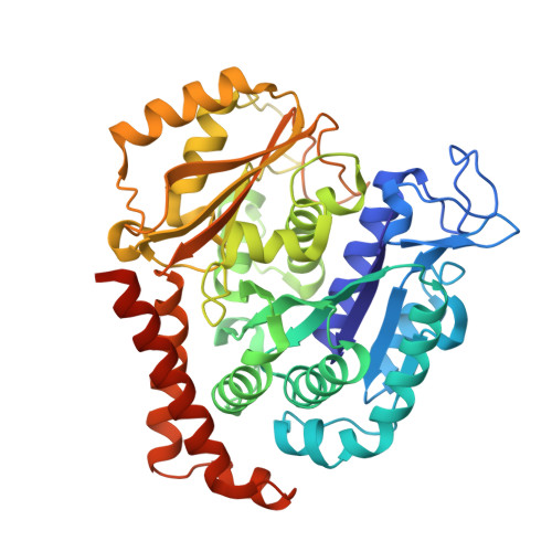
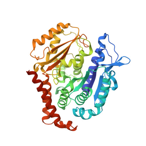
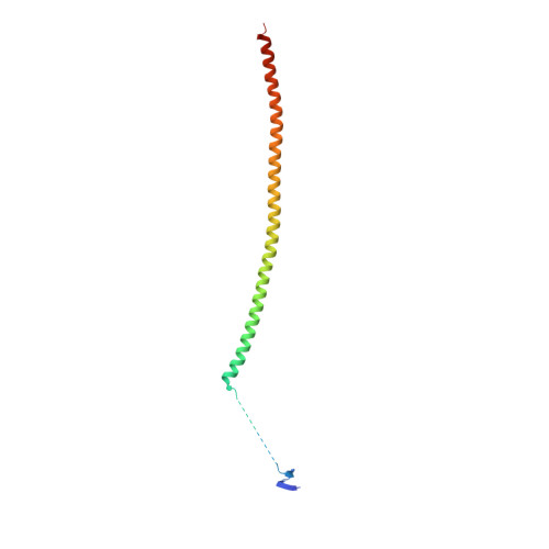
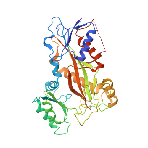
    Microtubules are dynamic assemblies of αβ-tubulin heterodimers and have been recognized as highly attractive targets for cancer chemotherapy. A broad range of agents bind to tubulin and interfere with microtubule assembly. Despite having a long history of characterization, colchicine binding site inhibitors (CBSIs) have not yet reached the commercial phase as anti-cancer drugs to date. We determined the structures of tubulin complexed with a set of structurally diverse CBSIs (lexibulin, nocodazole, plinabulin and tivantinib), among which nocodazole and tivantinib are both binary-function inhibitors targeting cancer-related kinases and microtubules simultaneously. High resolution structures revealed the detailed interactions between these ligands and tubulin. Our results showed that the binding modes of the CBSIs were different from previous docking models, highlighting the importance of crystal structure information in structure-based drug design. A real structure-based pharmacophore was proposed to rationalize key common interactions of the CBSIs at the colchicine domain. Our studies provide a solid structural basis for developing new anti-cancer agents for the colchicine binding site.


  • Organizational Affiliation

    State Key Laboratory of Biotherapy and Cancer Center, West China Hospital, Collaborative Innovation Center for Biotherapy, Sichuan University, Chengdu, China.


Macromolecules
Find similar proteins by:  (by identity cutoff)  |  3D Structure
Entity ID: 1
MoleculeChains Sequence LengthOrganismDetailsImage
Tubulin alpha
A, C
450Sus barbatusMutation(s): 0 
UniProt
Find proteins for A0A0R4I993 (Sus barbatus)
Explore A0A0R4I993 
Go to UniProtKB:  A0A0R4I993
Entity Groups  
Sequence Clusters30% Identity50% Identity70% Identity90% Identity95% Identity100% Identity
UniProt GroupA0A0R4I993
Sequence Annotations
Expand
  • Reference Sequence
Find similar proteins by:  (by identity cutoff)  |  3D Structure
Entity ID: 2
MoleculeChains Sequence LengthOrganismDetailsImage
Tubulin beta-2 chain
B, D
445Gallus gallusMutation(s): 0 
UniProt
Find proteins for P32882 (Gallus gallus)
Explore P32882 
Go to UniProtKB:  P32882
Entity Groups  
Sequence Clusters30% Identity50% Identity70% Identity90% Identity95% Identity100% Identity
UniProt GroupP32882
Sequence Annotations
Expand
  • Reference Sequence
Find similar proteins by:  (by identity cutoff)  |  3D Structure
Entity ID: 3
MoleculeChains Sequence LengthOrganismDetailsImage
Stathmin-4143Rattus norvegicusMutation(s): 0 
Gene Names: Stmn4
UniProt
Find proteins for P63043 (Rattus norvegicus)
Explore P63043 
Go to UniProtKB:  P63043
Entity Groups  
Sequence Clusters30% Identity50% Identity70% Identity90% Identity95% Identity100% Identity
UniProt GroupP63043
Sequence Annotations
Expand
  • Reference Sequence
Find similar proteins by:  (by identity cutoff)  |  3D Structure
Entity ID: 4
MoleculeChains Sequence LengthOrganismDetailsImage
Uncharacterized protein384Gallus gallusMutation(s): 0 
Gene Names: TTL
Entity Groups  
Sequence Clusters30% Identity50% Identity70% Identity90% Identity95% Identity100% Identity
Sequence Annotations
Expand
  • Reference Sequence
Small Molecules
Ligands 8 Unique
IDChains Name / Formula / InChI Key2D Diagram3D Interactions
GTP
Query on GTP

Download Ideal Coordinates CCD File 
G [auth A],
O [auth C]
GUANOSINE-5'-TRIPHOSPHATE
C10 H16 N5 O14 P3
XKMLYUALXHKNFT-UUOKFMHZSA-N
ACP
Query on ACP

Download Ideal Coordinates CCD File 
U [auth F]PHOSPHOMETHYLPHOSPHONIC ACID ADENYLATE ESTER
C11 H18 N5 O12 P3
UFZTZBNSLXELAL-IOSLPCCCSA-N
GDP
Query on GDP

Download Ideal Coordinates CCD File 
K [auth B],
R [auth D]
GUANOSINE-5'-DIPHOSPHATE
C10 H15 N5 O11 P2
QGWNDRXFNXRZMB-UUOKFMHZSA-N
NZO
Query on NZO

Download Ideal Coordinates CCD File 
N [auth B],
T [auth D]
nocodazole
C14 H11 N3 O3 S
KYRVNWMVYQXFEU-UHFFFAOYSA-N
MES
Query on MES

Download Ideal Coordinates CCD File 
M [auth B],
S [auth D]
2-(N-MORPHOLINO)-ETHANESULFONIC ACID
C6 H13 N O4 S
SXGZJKUKBWWHRA-UHFFFAOYSA-N
GOL
Query on GOL

Download Ideal Coordinates CCD File 
J [auth A]GLYCEROL
C3 H8 O3
PEDCQBHIVMGVHV-UHFFFAOYSA-N
CA
Query on CA

Download Ideal Coordinates CCD File 
I [auth A],
Q [auth C]
CALCIUM ION
Ca
BHPQYMZQTOCNFJ-UHFFFAOYSA-N
MG
Query on MG

Download Ideal Coordinates CCD File 
H [auth A],
L [auth B],
P [auth C]
MAGNESIUM ION
Mg
JLVVSXFLKOJNIY-UHFFFAOYSA-N
Binding Affinity Annotations 
IDSourceBinding Affinity
NZO BindingDB:  5CA1 IC50: min: 760, max: 5700 (nM) from 2 assay(s)
Experimental Data & Validation

Experimental Data

  • Method: X-RAY DIFFRACTION
  • Resolution: 2.40 Å
  • R-Value Free: 0.225 
  • R-Value Work: 0.192 
  • R-Value Observed: 0.193 
  • Space Group: P 21 21 21
Unit Cell:
Length ( Å )Angle ( ˚ )
a = 105.44α = 90
b = 158.362β = 90
c = 180.692γ = 90
Software Package:
Software NamePurpose
PHENIXrefinement
HKL-3000data reduction
HKL-3000data scaling
PHASERphasing

Structure Validation

View Full Validation Report


Ligand Structure Quality Assessment 


Entry History 

Deposition Data

Revision History  (Full details and data files)

  • Version 1.0: 2015-11-04
    Type: Initial release
  • Version 1.1: 2016-01-27
    Changes: Database references
  • Version 1.2: 2023-11-08
    Changes: Data collection, Database references, Derived calculations, Refinement description