5C5E

Structure of KaiA dimer in complex with C-terminal KaiC peptide at 2.8 A resolution


Experimental Data Snapshot

  • Method: X-RAY DIFFRACTION
  • Resolution: 2.82 Å
  • R-Value Free: 0.303 
  • R-Value Work: 0.240 
  • R-Value Observed: 0.243 

wwPDB Validation   3D Report Full Report


Ligand Structure Quality Assessment 


This is version 1.4 of the entry. See complete history


Literature

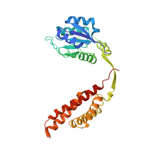
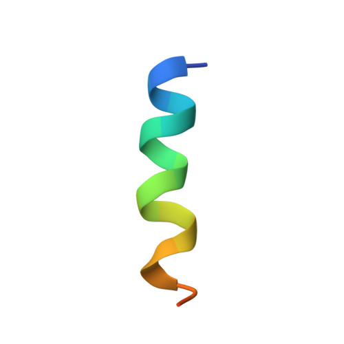
Protein-Protein Interactions in the Cyanobacterial Circadian Clock: Structure of KaiA Dimer in Complex with C-Terminal KaiC Peptides at 2.8 angstrom Resolution.

Pattanayek, R.Egli, M.

(2015) Biochemistry 54: 4575-4578

  • DOI: https://doi.org/10.1021/acs.biochem.5b00694
  • Primary Citation of Related Structures:  
    5C5E

  • PubMed Abstract: 

    In the cyanobacterial circadian clock, the KaiA, -B, and -C proteins with ATP constitute a post-translational oscillator. KaiA stimulates the KaiC autokinase, and KaiB antagonizes KaiA action. KaiA contacts the intrinsically disordered C-terminal regions of KaiC hexamer to promote phosphorylation across subunit interfaces. The crystal structure of KaiA dimer from Synechococcus elongatus with two KaiC C-terminal 20mer peptides bound reveals that the latter adopt an α-helical conformation and contact KaiA α-helical bundles via mostly hydrophobic interactions. This complex and the crystal structure of KaiC hexamer with truncated C-terminal tails can be fit into the electron microscopy (EM) density of the KaiA:KaiC complex. The hybrid model helps rationalize clock phenotypes of KaiA and KaiC mutants.


  • Organizational Affiliation

    Department of Biochemistry, Vanderbilt University, School of Medicine, Nashville, Tennessee 37232, United States.


Macromolecules
Find similar proteins by:  (by identity cutoff)  |  3D Structure
Entity ID: 1
MoleculeChains Sequence LengthOrganismDetailsImage
Circadian clock protein KaiAA,
C [auth B]
290Synechococcus elongatus PCC 7942 = FACHB-805Mutation(s): 0 
Gene Names: kaiASynpcc7942_1218see0009
UniProt
Find proteins for Q79PF6 (Synechococcus elongatus (strain ATCC 33912 / PCC 7942 / FACHB-805))
Explore Q79PF6 
Go to UniProtKB:  Q79PF6
Entity Groups  
Sequence Clusters30% Identity50% Identity70% Identity90% Identity95% Identity100% Identity
UniProt GroupQ79PF6
Sequence Annotations
Expand
  • Reference Sequence

Find similar proteins by:  Sequence   |   3D Structure  

Entity ID: 2
MoleculeChains Sequence LengthOrganismDetailsImage
KaiC C-terminal peptideB [auth G],
D [auth H]
20synthetic constructMutation(s): 0 
UniProt
Find proteins for Q79PF4 (Synechococcus elongatus (strain ATCC 33912 / PCC 7942 / FACHB-805))
Explore Q79PF4 
Go to UniProtKB:  Q79PF4
Entity Groups  
Sequence Clusters30% Identity50% Identity70% Identity90% Identity95% Identity100% Identity
UniProt GroupQ79PF4
Sequence Annotations
Expand
  • Reference Sequence
Small Molecules
Ligands 1 Unique
IDChains Name / Formula / InChI Key2D Diagram3D Interactions
52M
Query on 52M

Download Ideal Coordinates CCD File 
E [auth A],
F [auth B]
2-(6-hydroxy-3-oxo-3H-xanthen-9-yl)-5-[(sulfanylcarbonyl)amino]benzoic acid
C21 H13 N O6 S
YMGUTVDFHHFZIJ-UHFFFAOYSA-N
Experimental Data & Validation

Experimental Data

  • Method: X-RAY DIFFRACTION
  • Resolution: 2.82 Å
  • R-Value Free: 0.303 
  • R-Value Work: 0.240 
  • R-Value Observed: 0.243 
  • Space Group: P 43 21 2
Unit Cell:
Length ( Å )Angle ( ˚ )
a = 97.43α = 90
b = 97.43β = 90
c = 124.52γ = 90
Software Package:
Software NamePurpose
PHENIXrefinement
HKL-2000data reduction
PHASERphasing
Cootmodel building
Aimlessdata scaling

Structure Validation

View Full Validation Report


Ligand Structure Quality Assessment 


Entry History & Funding Information

Deposition Data


Funding OrganizationLocationGrant Number
National Institutes of Health/National Institute of General Medical Sciences (NIH/NIGMS)United StatesGM073845

Revision History  (Full details and data files)

  • Version 1.0: 2015-08-05
    Type: Initial release
  • Version 1.1: 2015-08-12
    Changes: Database references
  • Version 1.2: 2017-09-27
    Changes: Author supporting evidence, Database references, Derived calculations
  • Version 1.3: 2019-12-25
    Changes: Author supporting evidence
  • Version 1.4: 2023-09-27
    Changes: Data collection, Database references, Refinement description