5C25

Crystal Structure of HIV-1 Reverse Transcriptase in Complex with 6-((4-((4-cyanophenyl)amino)-1,3,5-triazin-2-yl)amino)-5,7-dimethyl-2-naphthonitrile (JLJ639), a Non-nucleoside Inhibitor


Experimental Data Snapshot

  • Method: X-RAY DIFFRACTION
  • Resolution: 2.84 Å
  • R-Value Free: 0.263 
  • R-Value Work: 0.216 
  • R-Value Observed: 0.218 

wwPDB Validation   3D Report Full Report


Ligand Structure Quality Assessment 


This is version 1.3 of the entry. See complete history


Literature

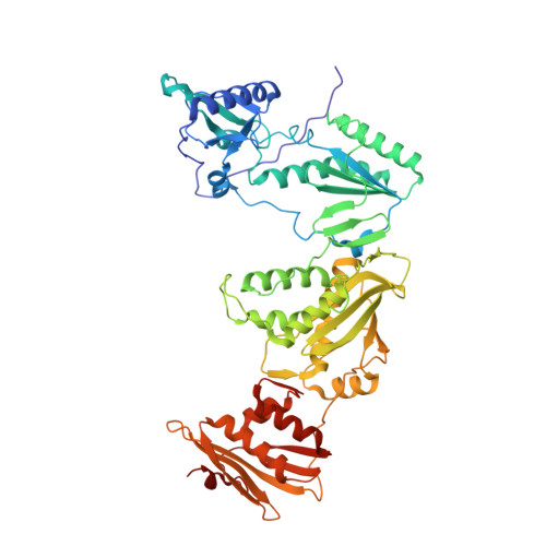
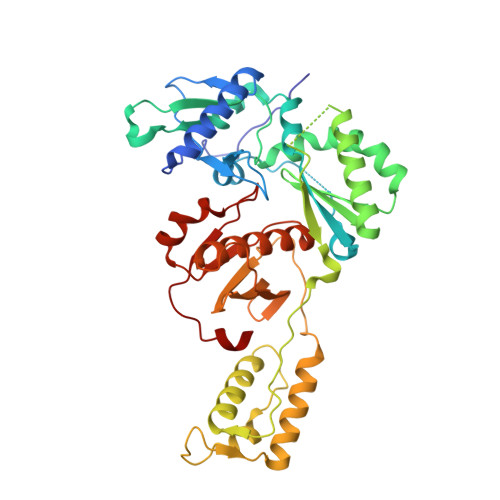
Discovery and crystallography of bicyclic arylaminoazines as potent inhibitors of HIV-1 reverse transcriptase.

Lee, W.G.Frey, K.M.Gallardo-Macias, R.Spasov, K.A.Chan, A.H.Anderson, K.S.Jorgensen, W.L.

(2015) Bioorg Med Chem Lett 25: 4824-4827

  • DOI: 10.1016/j.bmcl.2015.06.074
  • Primary Citation of Related Structures:  
    5C24, 5C25

  • PubMed Abstract: 
  • Non-nucleoside inhibitors of HIV-1 reverse transcriptase (HIV-RT) are reported that incorporate a 7-indolizinylamino or 2-naphthylamino substituent on a pyrimidine or 1,3,5-triazine core. The most potent compounds show below 10 nanomolar activity towards wild-type HIV-1 and variants bearing Tyr181Cys and Lys103Asn/Tyr181Cys resistance mutations ...

    Non-nucleoside inhibitors of HIV-1 reverse transcriptase (HIV-RT) are reported that incorporate a 7-indolizinylamino or 2-naphthylamino substituent on a pyrimidine or 1,3,5-triazine core. The most potent compounds show below 10 nanomolar activity towards wild-type HIV-1 and variants bearing Tyr181Cys and Lys103Asn/Tyr181Cys resistance mutations. The compounds also feature good aqueous solubility. Crystal structures for two complexes enhance the analysis of the structure-activity data.


    Organizational Affiliation

    Department of Chemistry, Yale University, New Haven, CT 06520-8107, USA. Electronic address: william.jorgensen@yale.edu.


Macromolecules
Find similar proteins by:  (by identity cutoff)  |  3D Structure
Entity ID: 1
MoleculeChainsSequence LengthOrganismDetailsImage
HIV-1 REVERSE TRANSCRIPTASE, P66 SUBUNITA557Human immunodeficiency virus type 1 BH10Mutation(s): 3 
Gene Names: gag-pol
EC: 3.4.23.16 (PDB Primary Data), 2.7.7.49 (PDB Primary Data), 2.7.7.7 (PDB Primary Data), 3.1.26.13 (PDB Primary Data), 3.1.13.2 (PDB Primary Data), 2.7.7 (UniProt), 3.1 (UniProt)
UniProt
Find proteins for P03366 (Human immunodeficiency virus type 1 group M subtype B (isolate BH10))
Explore P03366 
Go to UniProtKB:  P03366
Entity Groups  
Sequence Clusters30% Identity50% Identity70% Identity90% Identity95% Identity100% Identity
UniProt GroupP03366
Protein Feature View
Expand
  • Reference Sequence
Find similar proteins by:  (by identity cutoff)  |  3D Structure
Entity ID: 2
MoleculeChainsSequence LengthOrganismDetailsImage
HIV-1 REVERSE TRANSCRIPTASE, P51 SUBUNITB428Human immunodeficiency virus type 1 BH10Mutation(s): 1 
Gene Names: gag-pol
EC: 3.4.23.16 (PDB Primary Data), 2.7.7.49 (PDB Primary Data), 2.7.7.7 (PDB Primary Data), 3.1.26.13 (PDB Primary Data), 3.1.13.2 (PDB Primary Data), 2.7.7 (UniProt), 3.1 (UniProt)
UniProt
Find proteins for P03366 (Human immunodeficiency virus type 1 group M subtype B (isolate BH10))
Explore P03366 
Go to UniProtKB:  P03366
Entity Groups  
Sequence Clusters30% Identity50% Identity70% Identity90% Identity95% Identity100% Identity
UniProt GroupP03366
Protein Feature View
Expand
  • Reference Sequence
Small Molecules
Ligands 3 Unique
IDChainsName / Formula / InChI Key2D Diagram3D Interactions
639
Query on 639

Download Ideal Coordinates CCD File 
C [auth A]6-({4-[(4-cyanophenyl)amino]-1,3,5-triazin-2-yl}amino)-5,7- dimethyl-2-naphthonitrile
C23 H17 N7
KPKGAKOSWKZAAI-UHFFFAOYSA-N
 Ligand Interaction
SO4
Query on SO4

Download Ideal Coordinates CCD File 
D [auth A],
E [auth A]
SULFATE ION
O4 S
QAOWNCQODCNURD-UHFFFAOYSA-L
 Ligand Interaction
MG
Query on MG

Download Ideal Coordinates CCD File 
F [auth A]MAGNESIUM ION
Mg
JLVVSXFLKOJNIY-UHFFFAOYSA-N
 Ligand Interaction
Experimental Data & Validation

Experimental Data

  • Method: X-RAY DIFFRACTION
  • Resolution: 2.84 Å
  • R-Value Free: 0.263 
  • R-Value Work: 0.216 
  • R-Value Observed: 0.218 
  • Space Group: C 1 2 1
  • Diffraction Data DOI: 10.15785/SBGRID/168
Unit Cell:
Length ( Å )Angle ( ˚ )
a = 162.554α = 90
b = 72.969β = 100.66
c = 109.479γ = 90
Software Package:
Software NamePurpose
PHENIXrefinement
XDSdata reduction
XDSdata scaling
PHASERphasing

Structure Validation

View Full Validation Report


Ligand Structure Quality Assessment 


Entry History & Funding Information

Deposition Data


Funding OrganizationLocationGrant Number
National Institutes of Health/National Institute Of Allergy and Infectious Diseases (NIH/NIAID)United StatesAI27690
National Institutes of Health/National Institute Of Allergy and Infectious Diseases (NIH/NIAID)United StatesAI44616
National Institutes of Health/National Institute of General Medical Sciences (NIH/NIGMS)United StatesGM32136
National Institutes of Health/National Institute of General Medical Sciences (NIH/NIGMS)United StatesGM49551
National Institutes of Health/National Institute Of Allergy and Infectious Diseases (NIH/NIAID)United StatesAI104334

Revision History  (Full details and data files)

  • Version 1.0: 2015-07-29
    Type: Initial release
  • Version 1.1: 2015-10-28
    Changes: Database references
  • Version 1.2: 2017-09-13
    Changes: Author supporting evidence, Database references, Derived calculations
  • Version 1.3: 2019-12-11
    Changes: Author supporting evidence