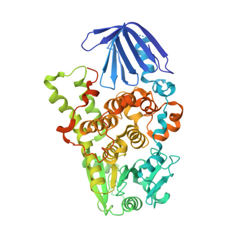
5C16

Myotubularin-related proetin 1


Experimental Data Snapshot

  • Method: X-RAY DIFFRACTION
  • Resolution: 2.07 Å
  • R-Value Free: 0.274 
  • R-Value Work: 0.220 
  • R-Value Observed: 0.223 

wwPDB Validation   3D Report Full Report


This is version 1.1 of the entry. See complete history


Literature

Crystal Structure of Human Myotubularin-Related Protein 1 Provides Insight into the Structural Basis of Substrate Specificity

Bong, S.M.Son, K.B.Yang, S.W.Park, J.W.Cho, J.W.Kim, K.T.Kim, H.Kim, S.J.Kim, Y.J.Lee, B.I.

(2016) PLoS One 11: e0152611-e0152611

  • DOI: https://doi.org/10.1371/journal.pone.0152611
  • Primary Citation of Related Structures:  
    5C16

  • PubMed Abstract: 

    Myotubularin-related protein 1 (MTMR1) is a phosphatase that belongs to the tyrosine/dual-specificity phosphatase superfamily. MTMR1 has been shown to use phosphatidylinositol 3-monophosphate (PI(3)P) and/or phosphatidylinositol 3,5-bisphosphate (PI(3,5)P2) as substrates. Here, we determined the crystal structure of human MTMR1. The refined model consists of the Pleckstrin homology (PH)-GRAM and phosphatase (PTP) domains. The overall structure was highly similar to the previously reported MTMR2 structure. Interestingly, two phosphate molecules were coordinated by strictly conserved residues located in the C(X)5R motif of the active site. Additionally, our biochemical studies confirmed the substrate specificity of MTMR1 for PI(3)P and PI(3,5)P2 over other phosphatidylinositol phosphates. Our structural and enzymatic analyses provide insight into the catalytic mechanism and biochemical properties of MTMR1.


  • Organizational Affiliation

    Research institute, National Cancer Center, Goyang, Gyeonggi 10408, Republic of Korea.


Macromolecules
Find similar proteins by:  (by identity cutoff)  |  3D Structure
Entity ID: 1
MoleculeChains Sequence LengthOrganismDetailsImage
Myotubularin-related protein 1
A, B, C, D
571Homo sapiensMutation(s): 1 
Gene Names: MTMR1
EC: 3.1.3.64
Membrane Entity: Yes 
UniProt & NIH Common Fund Data Resources
Find proteins for Q13613 (Homo sapiens)
Explore Q13613 
Go to UniProtKB:  Q13613
PHAROS:  Q13613
GTEx:  ENSG00000063601 
Entity Groups  
Sequence Clusters30% Identity50% Identity70% Identity90% Identity95% Identity100% Identity
UniProt GroupQ13613
Sequence Annotations
Expand
  • Reference Sequence
Experimental Data & Validation

Experimental Data

  • Method: X-RAY DIFFRACTION
  • Resolution: 2.07 Å
  • R-Value Free: 0.274 
  • R-Value Work: 0.220 
  • R-Value Observed: 0.223 
  • Space Group: P 1
Unit Cell:
Length ( Å )Angle ( ˚ )
a = 67.219α = 87.6
b = 96.587β = 86.07
c = 97.581γ = 77.33
Software Package:
Software NamePurpose
PHENIXrefinement
Cootmodel building
HKL-2000data scaling
HKL-2000data processing

Structure Validation

View Full Validation Report


Entry History 

Deposition Data

Revision History  (Full details and data files)

  • Version 1.0: 2016-04-27
    Type: Initial release
  • Version 1.1: 2024-03-20
    Changes: Data collection, Database references, Derived calculations