5C15

K428A mutant nuclease domain of the large terminase subunit gp2 of bacterial virus Sf6 with Manganese


Experimental Data Snapshot

  • Method: X-RAY DIFFRACTION
  • Resolution: 1.57 Å
  • R-Value Free: 0.186 
  • R-Value Work: 0.164 
  • R-Value Observed: 0.165 

wwPDB Validation   3D Report Full Report


This is version 1.5 of the entry. See complete history


Literature

Two distinct modes of metal ion binding in the nuclease active site of a viral DNA-packaging terminase: insight into the two-metal-ion catalytic mechanism.

Zhao, H.Lin, Z.Lynn, A.Y.Varnado, B.Beutler, J.A.Murelli, R.P.Le Grice, S.F.Tang, L.

(2015) Nucleic Acids Res 43: 11003-11016

  • DOI: https://doi.org/10.1093/nar/gkv1018
  • Primary Citation of Related Structures:  
    5C10, 5C12, 5C15, 5C2D, 5C2F

  • PubMed Abstract: 

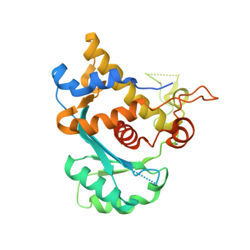
    Many dsDNA viruses encode DNA-packaging terminases, each containing a nuclease domain that resolves concatemeric DNA into genome-length units. Terminase nucleases resemble the RNase H-superfamily nucleotidyltransferases in folds, and share a two-metal-ion catalytic mechanism. Here we show that residue K428 of a bacteriophage terminase gp2 nuclease domain mediates binding of the metal cofactor Mg(2+). A K428A mutation allows visualization, at high resolution, of a metal ion binding mode with a coupled-octahedral configuration at the active site, exhibiting an unusually short metal-metal distance of 2.42 Å. Such proximity of the two metal ions may play an essential role in catalysis by generating a highly positive electrostatic niche to enable formation of the negatively charged pentacovalent phosphate transition state, and provides the structural basis for distinguishing Mg(2+) from Ca(2+). Using a metal ion chelator β-thujaplicinol as a molecular probe, we observed a second mode of metal ion binding at the active site, mimicking the DNA binding state. Arrangement of the active site residues differs drastically from those in RNase H-like nucleases, suggesting a drifting of the active site configuration during evolution. The two distinct metal ion binding modes unveiled mechanistic details of the two-metal-ion catalysis at atomic resolution.


  • Organizational Affiliation

    Department of Molecular Biosciences, University of Kansas, 1200 Sunnyside Avenue, Lawrence, KS 66045, USA.


Macromolecules
Find similar proteins by:  (by identity cutoff)  |  3D Structure
Entity ID: 1
MoleculeChains Sequence LengthOrganismDetailsImage
Gene 2 protein278Lederbergvirus Sf6Mutation(s): 3 
UniProt
Find proteins for Q716H3 (Shigella phage Sf6)
Explore Q716H3 
Go to UniProtKB:  Q716H3
Entity Groups  
Sequence Clusters30% Identity50% Identity70% Identity90% Identity95% Identity100% Identity
UniProt GroupQ716H3
Sequence Annotations
Expand
  • Reference Sequence
Experimental Data & Validation

Experimental Data

  • Method: X-RAY DIFFRACTION
  • Resolution: 1.57 Å
  • R-Value Free: 0.186 
  • R-Value Work: 0.164 
  • R-Value Observed: 0.165 
  • Space Group: C 1 2 1
Unit Cell:
Length ( Å )Angle ( ˚ )
a = 132.766α = 90
b = 57.365β = 98.71
c = 46.646γ = 90
Software Package:
Software NamePurpose
PHENIXrefinement
HKL-2000data collection
PDB_EXTRACTdata extraction
HKL-2000data reduction
HKL-2000data scaling
PHENIXphasing

Structure Validation

View Full Validation Report


Entry History & Funding Information

Deposition Data


Funding OrganizationLocationGrant Number
National Institutes of Health/National Institute of General Medical Sciences (NIH/NIGMS)United StatesR01GM090010

Revision History  (Full details and data files)

  • Version 1.0: 2015-10-21
    Type: Initial release
  • Version 1.1: 2015-12-30
    Changes: Database references
  • Version 1.2: 2017-09-06
    Changes: Author supporting evidence, Database references, Derived calculations
  • Version 1.3: 2017-09-27
    Changes: Data collection
  • Version 1.4: 2019-12-25
    Changes: Author supporting evidence
  • Version 1.5: 2023-11-15
    Changes: Data collection, Database references, Derived calculations, Structure summary