5C14

Crystal structure of PECAM-1 D1D2 domain


Experimental Data Snapshot

  • Method: X-RAY DIFFRACTION
  • Resolution: 2.80 Å
  • R-Value Free: 0.279 
  • R-Value Work: 0.238 
  • R-Value Observed: 0.239 

wwPDB Validation   3D Report Full Report


Ligand Structure Quality Assessment 


This is version 1.2 of the entry. See complete history


Literature

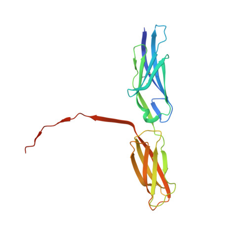
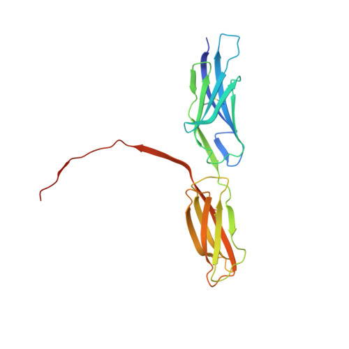
Structural basis for PECAM-1 homophilic binding.

Paddock, C.Zhou, D.Lertkiatmongkol, P.Newman, P.J.Zhu, J.

(2016) Blood 127: 1052-1061

  • DOI: https://doi.org/10.1182/blood-2015-07-660092
  • Primary Citation of Related Structures:  
    5C14

  • PubMed Abstract: 

    Platelet endothelial cell adhesion molecule-1 (PECAM-1) is a 130-kDa member of the immunoglobulin gene superfamily (IgSF) that is present on the surface of circulating platelets and leukocytes, and highly expressed at the junctions of confluent endothelial cell monolayers. PECAM-1-mediated homophilic interactions, known to be mediated by its 2 amino-terminal immunoglobulin homology domains, are essential for concentrating PECAM-1 at endothelial cell intercellular junctions, where it functions to facilitate diapedesis, maintain vascular integrity, and transmit survival signals into the cell. Given the importance of PECAM-1-mediated homophilic interactions in mediating each of these cell physiological events, and to reveal the nature and orientation of the PECAM-1-PECAM-1 homophilic-binding interface, we undertook studies aimed at determining the crystal structure of the PECAM-1 homophilic-binding domain, which is composed of amino-terminal immunoglobulin homology domains 1 and 2 (IgD1 and IgD2). The crystal structure revealed that both IgD1 and IgD2 exhibit a classical IgSF fold, having a β-sandwich topology formed by 2 sheets of antiparallel β strands stabilized by the hallmark disulfide bond between the B and F strands. Interestingly, despite previous assignment to the C2 class of immunoglobulin-like domains, the structure of IgD1 reveals that it actually belongs to the I2 set of IgSF folds. Both IgD1 and IgD2 participate importantly in the formation of the trans homophilic-binding interface, with a total buried interface area of >2300 Å(2). These and other unique structural features of PECAM-1 allow for the development of an atomic-level model of the interactions that PECAM-1 forms during assembly of endothelial cell intercellular junctions.


  • Organizational Affiliation

    Blood Research Institute, BloodCenter of Wisconsin, Milwaukee, WI; and.


Macromolecules
Find similar proteins by:  (by identity cutoff)  |  3D Structure
Entity ID: 1
MoleculeChains Sequence LengthOrganismDetailsImage
Platelet endothelial cell adhesion molecule212Homo sapiensMutation(s): 0 
Gene Names: PECAM1
UniProt & NIH Common Fund Data Resources
Find proteins for P16284 (Homo sapiens)
Explore P16284 
Go to UniProtKB:  P16284
PHAROS:  P16284
GTEx:  ENSG00000261371 
Entity Groups  
Sequence Clusters30% Identity50% Identity70% Identity90% Identity95% Identity100% Identity
UniProt GroupP16284
Sequence Annotations
Expand
  • Reference Sequence
Find similar proteins by:  (by identity cutoff)  |  3D Structure
Entity ID: 2
MoleculeChains Sequence LengthOrganismDetailsImage
Platelet endothelial cell adhesion molecule212Homo sapiensMutation(s): 0 
Gene Names: PECAM1
UniProt & NIH Common Fund Data Resources
Find proteins for P16284 (Homo sapiens)
Explore P16284 
Go to UniProtKB:  P16284
PHAROS:  P16284
GTEx:  ENSG00000261371 
Entity Groups  
Sequence Clusters30% Identity50% Identity70% Identity90% Identity95% Identity100% Identity
UniProt GroupP16284
Sequence Annotations
Expand
  • Reference Sequence
Small Molecules
Ligands 4 Unique
IDChains Name / Formula / InChI Key2D Diagram3D Interactions
NAG
Query on NAG

Download Ideal Coordinates CCD File 
G [auth A],
H [auth A],
I [auth A],
M [auth B],
N [auth B]
2-acetamido-2-deoxy-beta-D-glucopyranose
C8 H15 N O6
OVRNDRQMDRJTHS-FMDGEEDCSA-N
TRS
Query on TRS

Download Ideal Coordinates CCD File 
J [auth A]2-AMINO-2-HYDROXYMETHYL-PROPANE-1,3-DIOL
C4 H12 N O3
LENZDBCJOHFCAS-UHFFFAOYSA-O
GOL
Query on GOL

Download Ideal Coordinates CCD File 
C [auth A],
D [auth A],
E [auth A],
K [auth B],
L [auth B]
GLYCEROL
C3 H8 O3
PEDCQBHIVMGVHV-UHFFFAOYSA-N
CU
Query on CU

Download Ideal Coordinates CCD File 
F [auth A]COPPER (II) ION
Cu
JPVYNHNXODAKFH-UHFFFAOYSA-N
Modified Residues  1 Unique
IDChains TypeFormula2D DiagramParent
MSE
Query on MSE
A
L-PEPTIDE LINKINGC5 H11 N O2 SeMET
Experimental Data & Validation

Experimental Data

  • Method: X-RAY DIFFRACTION
  • Resolution: 2.80 Å
  • R-Value Free: 0.279 
  • R-Value Work: 0.238 
  • R-Value Observed: 0.239 
  • Space Group: I 41 2 2
Unit Cell:
Length ( Å )Angle ( ˚ )
a = 104.01α = 90
b = 104.01β = 90
c = 281.834γ = 90
Software Package:
Software NamePurpose
PHENIXrefinement
XDSdata reduction
SCALAdata scaling
PHENIXphasing

Structure Validation

View Full Validation Report


Ligand Structure Quality Assessment 


Entry History 

Deposition Data

Revision History  (Full details and data files)

  • Version 1.0: 2016-01-06
    Type: Initial release
  • Version 1.1: 2016-03-09
    Changes: Database references
  • Version 1.2: 2020-07-29
    Type: Remediation
    Reason: Carbohydrate remediation
    Changes: Data collection, Derived calculations, Structure summary