5C03

Crystal Structure of kinase


Experimental Data Snapshot

  • Method: X-RAY DIFFRACTION
  • Resolution: 1.90 Å
  • R-Value Free: 0.218 
  • R-Value Work: 0.181 
  • R-Value Observed: 0.183 

wwPDB Validation   3D Report Full Report


Ligand Structure Quality Assessment 


This is version 1.3 of the entry. See complete history


Literature

Structural and Functional Characterization of the JH2 Pseudokinase Domain of JAK Family Tyrosine Kinase 2 (TYK2).

Min, X.Ungureanu, D.Maxwell, S.Hammaren, H.Thibault, S.Hillert, E.K.Ayres, M.Greenfield, B.Eksterowicz, J.Gabel, C.Walker, N.Silvennoinen, O.Wang, Z.

(2015) J Biol Chem 290: 27261-27270

  • DOI: https://doi.org/10.1074/jbc.M115.672048
  • Primary Citation of Related Structures:  
    5C01, 5C03

  • PubMed Abstract: 

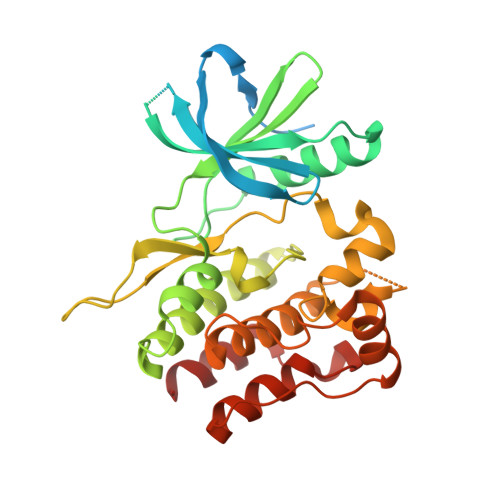
    JAK (Janus family of cytoplasmic tyrosine kinases) family tyrosine kinase 2 (TYK2) participates in signaling through cytokine receptors involved in immune responses and inflammation. JAKs are characterized by dual kinase domain: a tyrosine kinase domain (JH1) that is preceded by a pseudokinase domain (JH2). The majority of disease-associated mutations in JAKs map to JH2, demonstrating its central regulatory function. JH2s were considered catalytically inactive, but JAK2 JH2 was found to have low autoregulatory catalytic activity. Whether the other JAK JH2s share ATP binding and enzymatic activity has been unclear. Here we report the crystal structure of TYK2 JH2 in complex with adenosine 5'-O-(thiotriphosphate) (ATP-γS) and characterize its nucleotide binding by biochemical and biophysical methods. TYK2 JH2 did not show phosphotransfer activity, but it binds ATP and the nucleotide binding stabilizes the protein without inducing major conformational changes. Mutation of the JH2 ATP-binding pocket increased basal TYK2 phosphorylation and downstream signaling. The overall structural characteristics of TYK2 JH2 resemble JAK2 JH2, but distinct stabilizing molecular interactions around helix αAL in the activation loop provide a structural basis for differences in substrate access and catalytic activities among JAK family JH2s. The structural and biochemical data suggest that ATP binding is functionally important for both TYK2 and JAK2 JH2s, whereas the regulatory phosphorylation appears to be a unique property of JAK2. Finally, the co-crystal structure of TYK2 JH2 complexed with a small molecule inhibitor demonstrates that JH2 is accessible to ATP-competitive compounds, which offers novel approaches for targeting cytokine signaling as well as potential therapeutic applications.


  • Organizational Affiliation

    Departments of Therapeutic Discovery, Amgen Inc., South San Francisco, California 94080.


Macromolecules
Find similar proteins by:  (by identity cutoff)  |  3D Structure
Entity ID: 1
MoleculeChains Sequence LengthOrganismDetailsImage
Non-receptor tyrosine-protein kinase TYK2
A, B
342Homo sapiensMutation(s): 0 
Gene Names: TYK2
EC: 2.7.10.2
UniProt & NIH Common Fund Data Resources
Find proteins for P29597 (Homo sapiens)
Explore P29597 
Go to UniProtKB:  P29597
PHAROS:  P29597
GTEx:  ENSG00000105397 
Entity Groups  
Sequence Clusters30% Identity50% Identity70% Identity90% Identity95% Identity100% Identity
UniProt GroupP29597
Sequence Annotations
Expand
  • Reference Sequence
Small Molecules
Ligands 4 Unique
IDChains Name / Formula / InChI Key2D Diagram3D Interactions
AGS
Query on AGS

Download Ideal Coordinates CCD File 
C [auth A],
K [auth B]
PHOSPHOTHIOPHOSPHORIC ACID-ADENYLATE ESTER
C10 H16 N5 O12 P3 S
NLTUCYMLOPLUHL-KQYNXXCUSA-N
GOL
Query on GOL

Download Ideal Coordinates CCD File 
E [auth A],
H [auth A],
I [auth A],
M [auth B],
Q [auth B]
GLYCEROL
C3 H8 O3
PEDCQBHIVMGVHV-UHFFFAOYSA-N
EDO
Query on EDO

Download Ideal Coordinates CCD File 
F [auth A]
G [auth A]
J [auth A]
N [auth B]
O [auth B]
F [auth A],
G [auth A],
J [auth A],
N [auth B],
O [auth B],
P [auth B]
1,2-ETHANEDIOL
C2 H6 O2
LYCAIKOWRPUZTN-UHFFFAOYSA-N
MG
Query on MG

Download Ideal Coordinates CCD File 
D [auth A],
L [auth B]
MAGNESIUM ION
Mg
JLVVSXFLKOJNIY-UHFFFAOYSA-N
Binding Affinity Annotations 
IDSourceBinding Affinity
AGS Binding MOAD:  5C03 Kd: 1.50e+4 (nM) from 1 assay(s)
Experimental Data & Validation

Experimental Data

  • Method: X-RAY DIFFRACTION
  • Resolution: 1.90 Å
  • R-Value Free: 0.218 
  • R-Value Work: 0.181 
  • R-Value Observed: 0.183 
  • Space Group: P 1 21 1
Unit Cell:
Length ( Å )Angle ( ˚ )
a = 56.12α = 90
b = 47.69β = 93.72
c = 112.75γ = 90
Software Package:
Software NamePurpose
REFMACrefinement
PDB_EXTRACTdata extraction
SCALAdata scaling
PHASERphasing
MOSFLMdata reduction

Structure Validation

View Full Validation Report


Ligand Structure Quality Assessment 


Entry History 

Deposition Data

Revision History  (Full details and data files)

  • Version 1.0: 2015-09-16
    Type: Initial release
  • Version 1.1: 2015-09-23
    Changes: Database references
  • Version 1.2: 2015-11-18
    Changes: Database references
  • Version 1.3: 2024-03-06
    Changes: Data collection, Database references, Derived calculations