5C02

Influenza A M2 transmembrane domain drug-resistant S31N mutant at pH 8.0


Experimental Data Snapshot

  • Method: X-RAY DIFFRACTION
  • Resolution: 1.59 Å
  • R-Value Free: 0.197 
  • R-Value Work: 0.160 
  • R-Value Observed: 0.163 

wwPDB Validation   3D Report Full Report


Ligand Structure Quality Assessment 


This is version 1.4 of the entry. See complete history


Literature

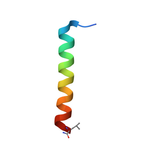
Crystal structure of the drug-resistant S31N influenza M2 proton channel.

Thomaston, J.L.DeGrado, W.F.

(2016) Protein Sci 25: 1551-1554

  • DOI: https://doi.org/10.1002/pro.2937
  • Primary Citation of Related Structures:  
    5C02

  • PubMed Abstract: 

    The M2 protein is a small proton channel found in the influenza A virus that is necessary for viral replication. The M2 channel is the target of a class of drugs called the adamantanes, which block the channel pore and prevent the virus from replicating. In recent decades mutations have arisen in M2 that prevent the adamantanes from binding to the channel pore, with the most prevalent of these mutations being S31N. Here we report the first crystal structure of the S31N mutant crystallized using lipidic cubic phase crystallization techniques and solved to 1.59 Å resolution. The Asn31 residues point directly into the center of the channel pore and form a hydrogen-bonded network that disrupts the drug-binding site. Ordered waters in the channel pore form a continuous hydrogen bonding network from Gly34 to His37.


  • Organizational Affiliation

    Department of Pharmaceutical Chemistry, University of California San Francisco, 555 Mission Bay Blvd. (S) Room 452V, San Francisco, California, 94158.


Macromolecules
Find similar proteins by:  (by identity cutoff)  |  3D Structure
Entity ID: 1
MoleculeChains Sequence LengthOrganismDetailsImage
Matrix protein 227Influenza A virus (A/Udorn/307/1972(H3N2))Mutation(s): 2 
Membrane Entity: Yes 
UniProt
Find proteins for P0DOF5 (Influenza A virus (strain A/Udorn/307/1972 H3N2))
Explore P0DOF5 
Go to UniProtKB:  P0DOF5
Entity Groups  
Sequence Clusters30% Identity50% Identity70% Identity90% Identity95% Identity100% Identity
UniProt GroupP0DOF5
Sequence Annotations
Expand
  • Reference Sequence
Experimental Data & Validation

Experimental Data

  • Method: X-RAY DIFFRACTION
  • Resolution: 1.59 Å
  • R-Value Free: 0.197 
  • R-Value Work: 0.160 
  • R-Value Observed: 0.163 
  • Space Group: I 4
Unit Cell:
Length ( Å )Angle ( ˚ )
a = 28.65α = 90
b = 28.65β = 90
c = 68.38γ = 90
Software Package:
Software NamePurpose
PHENIXrefinement
MOSFLMdata reduction
Aimlessdata scaling
PHASERphasing

Structure Validation

View Full Validation Report


Ligand Structure Quality Assessment 


Entry History & Funding Information

Deposition Data


Funding OrganizationLocationGrant Number
National Institutes of Health/National Institute of General Medical Sciences (NIH/NIGMS)United StatesR01-GM056423

Revision History  (Full details and data files)

  • Version 1.0: 2016-05-04
    Type: Initial release
  • Version 1.1: 2016-07-27
    Changes: Database references
  • Version 1.2: 2017-09-27
    Changes: Author supporting evidence, Database references, Derived calculations
  • Version 1.3: 2019-12-25
    Changes: Author supporting evidence
  • Version 1.4: 2023-09-27
    Changes: Data collection, Database references, Derived calculations, Refinement description