5BYK | pdb_00005byk

Schistosoma mansoni (Blood Fluke) Sulfotransferase/S-oxamniquine Complex


Experimental Data Snapshot

  • Method: X-RAY DIFFRACTION
  • Resolution: 1.28 Å
  • R-Value Free: 
    0.171 (Depositor), 0.174 (DCC) 
  • R-Value Work: 
    0.152 (Depositor), 0.155 (DCC) 
  • R-Value Observed: 
    0.152 (Depositor) 

Starting Model: experimental
View more details

wwPDB Validation 3D Report Full Report

Validation slider image for 5BYK

Ligand Structure Quality Assessment 


This is version 1.3 of the entry. See complete history

Literature

Structural and Functional Characterization of the Enantiomers of the Antischistosomal Drug Oxamniquine.

Taylor, A.B.Pica-Mattoccia, L.Polcaro, C.M.Donati, E.Cao, X.Basso, A.Guidi, A.Rugel, A.R.Holloway, S.P.Anderson, T.J.Hart, P.J.Cioli, D.LoVerde, P.T.

(2015) PLoS Negl Trop Dis 9: e0004132-e0004132

  • DOI: https://doi.org/10.1371/journal.pntd.0004132
  • Primary Citation Related Structures: 
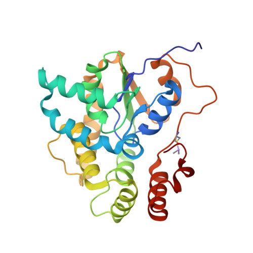
    5BYJ, 5BYK

  • PubMed Abstract: 

    For over two decades, a racemic mixture of oxamniquine (OXA) was administered to patients infected by Schistosoma mansoni, but whether one or both enantiomers exert antischistosomal activity was unknown. Recently, a ~30 kDa S. mansoni sulfotransferase (SmSULT) was identified as the target of OXA action. Here, we separate the OXA enantiomers using chromatographic methods and assign their optical activities as dextrorotary [(+)-OXA] or levorotary [(-)-OXA]. Crystal structures of the parasite enzyme in complex with optically pure (+)-OXA and (-)-OXA) reveal their absolute configurations as S- and R-, respectively. When tested in vitro, S-OXA demonstrated the bulk of schistosomicidal activity, while R-OXA had antischistosomal effects when present at relatively high concentrations. Crystal structures R-OXA•SmSULT and S-OXA•SmSULT complexes reveal similarities in the modes of OXA binding, but only the S-OXA enantiomer is observed in the structure of the enzyme exposed to racemic OXA. Together the data suggest the higher schistosomicidal activity of S-OXA is correlated with its ability to outcompete R-OXA binding the sulfotransferase active site. These findings have important implications for the design, syntheses, and dosing of new OXA-based antischistosomal compounds.


  • Organizational Affiliation
    • Departments of Biochemistry, the University of Texas Health Science Center, San Antonio, Texas, United States of America; X-ray Crystallography Core Laboratory, the University of Texas Health Science Center, San Antonio, Texas, United States of America.

Macromolecule Content 

  • Total Structure Weight: 30.81 kDa 
  • Atom Count: 2,491 
  • Modeled Residue Count: 254 
  • Deposited Residue Count: 259 
  • Unique protein chains: 1

Macromolecules

Find similar proteins by:|  3D Structure
Entity ID: 1
MoleculeChains  Sequence LengthOrganismDetailsImage
Sulfotransferase259Schistosoma mansoniMutation(s): 0 
Gene Names: Smp_089320
UniProt
Find proteins for G4VLE5 (Schistosoma mansoni)
Explore G4VLE5 
Go to UniProtKB:  G4VLE5
Entity Groups
Sequence Clusters30% Identity50% Identity70% Identity90% Identity95% Identity100% Identity
UniProt GroupG4VLE5
Sequence Annotations
Expand
Reference Sequence

Small Molecules

Ligands 3 Unique
IDChains Name / Formula / InChI Key2D Diagram3D Interactions
A3P

Query on A3P


Download:Ideal Coordinates CCD File
B [auth A]ADENOSINE-3'-5'-DIPHOSPHATE
C10 H15 N5 O10 P2
WHTCPDAXWFLDIH-KQYNXXCUSA-N
OAQ

Query on OAQ


Download:Ideal Coordinates CCD File
C [auth A]{(2S)-7-nitro-2-[(propan-2-ylamino)methyl]-1,2,3,4-tetrahydroquinolin-6-yl}methanol
C14 H21 N3 O3
XCGYUJZMCCFSRP-LBPRGKRZSA-N
NA

Query on NA


Download:Ideal Coordinates CCD File
D [auth A]SODIUM ION
Na
FKNQFGJONOIPTF-UHFFFAOYSA-N
Modified Residues  1 Unique
IDChains TypeFormula2D DiagramParent
CAS
Query on CAS
A
L-PEPTIDE LINKINGC5 H12 As N O2 SCYS

Experimental Data & Validation

Experimental Data

  • Method: X-RAY DIFFRACTION
  • Resolution: 1.28 Å
  • R-Value Free:  0.171 (Depositor), 0.174 (DCC) 
  • R-Value Work:  0.152 (Depositor), 0.155 (DCC) 
  • R-Value Observed: 0.152 (Depositor) 
Space Group: P 21 21 2
Unit Cell:
Length ( Å )Angle ( ˚ )
a = 140.587α = 90
b = 39.433β = 90
c = 54.003γ = 90
Software Package:
Software NamePurpose
PHENIXrefinement
XDSdata reduction
XDSdata scaling

Structure Validation

View Full Validation Report


Ligand Structure Quality Assessment 


Entry History 

& Funding Information

Deposition Data


Funding OrganizationLocationGrant Number
Welch FoundationUnited StatesAQ-1399

Revision History  (Full details and data files)

  • Version 1.0: 2015-09-30
    Type: Initial release
  • Version 1.1: 2015-11-18
    Changes: Database references, Structure summary
  • Version 1.2: 2023-09-27
    Changes: Data collection, Database references, Derived calculations, Refinement description
  • Version 1.3: 2024-10-16
    Changes: Structure summary