5BXQ | pdb_00005bxq

Structure of the NTF2:RanGDP complex


Experimental Data Snapshot

  • Method: X-RAY DIFFRACTION
  • Resolution: 2.50 Å
  • R-Value Free: 
    0.225 (Depositor), 0.228 (DCC) 
  • R-Value Work: 
    0.183 (Depositor), 0.192 (DCC) 
  • R-Value Observed: 
    0.186 (Depositor) 

wwPDB Validation 3D Report Full Report

Validation slider image for 5BXQ

Ligand Structure Quality Assessment 


This is version 1.2 of the entry. See complete history

Re-refinement Note

This entry reflects an alternative modeling of the original data in: 1A2K

Literature

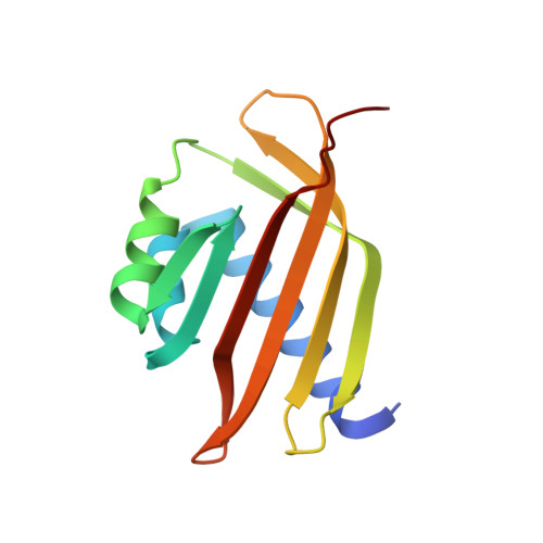
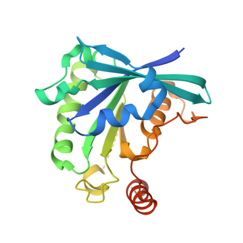
Structural basis for molecular recognition between nuclear transport factor 2 (NTF2) and the GDP-bound form of the Ras-family GTPase Ran.

Stewart, M.Kent, H.M.McCoy, A.J.

(1998) J Mol Biology 277: 635-646

  • DOI: https://doi.org/10.1006/jmbi.1997.1602
  • Primary Citation Related Structures: 
    5BXQ

  • PubMed Abstract: 

    Nuclear transport factor 2 (NTF2) and the Ras-family GTPase Ran are two soluble components of the nuclear protein import machinery. NTF2 binds GDP-Ran selectively and this interaction is important for efficient nuclear protein import in vivo. We have used X-ray crystallography to determine the structure of the macromolecular complex formed between GDP-Ran and nuclear transport factor 2 (NTF2) at 2.5 A resolution. The interaction interface involves primarily the putative switch II loop of Ran (residues 65 to 78) and the hydrophobic cavity and surrounding surface of NTF2. The major contribution to the interaction made by the switch II loop accounts for the ability of NTF2 to discriminate between GDP and GTP-bound forms of Ran. The aromatic side-chain of Ran Phe72 inserts into the NTF2 cavity and accounts for 22% of the surface area buried by the interaction interface, while salt bridges are formed between Lys71 and Arg76 of Ran with Asp92/Asp94 and Glu42 of NTF2, respectively. These salt bridges account for the inhibition of the Ran-NTF2 interaction by NTF2 mutants such as E42 K and D92/94N in which the negatively charged residues surrounding the cavity were altered. Because the interaction interface maintains the positions of key Ran residues involved in binding MgGDP, NTF2 binding may help stabilize the switch state of Ran, possibly in the context of targeting it to other components of the nuclear protein import machinery to specify directionality of transport. The binding of GDP-Ran at the NTF2 cavity raises the possibility that this interaction might be modulated by a metabolite or small molecule substrate for NTF2's putative enzymatic activity.


  • Organizational Affiliation
    • MRC Laboratory of Molecular Biology, Hills Road, Cambridge, CB2 2QH, England.

Macromolecule Content 

  • Total Structure Weight: 103.85 kDa 
  • Atom Count: 7,174 
  • Modeled Residue Count: 841 
  • Deposited Residue Count: 902 
  • Unique protein chains: 2

Macromolecules

Find similar proteins by:|  3D Structure
Entity ID: 1
MoleculeChains  Sequence LengthOrganismDetailsImage
Nuclear transport factor 2
A, B
127Rattus norvegicusMutation(s): 0 
Gene Names: Nutf2Ntf2
UniProt
Find proteins for P61972 (Rattus norvegicus)
Explore P61972 
Go to UniProtKB:  P61972
Entity Groups
Sequence Clusters30% Identity50% Identity70% Identity90% Identity95% Identity100% Identity
UniProt GroupP61972
Sequence Annotations
Expand
Reference Sequence
Find similar proteins by:|  3D Structure
Entity ID: 2
MoleculeChains  Sequence LengthOrganismDetailsImage
GTP-binding nuclear protein Ran
C, D, E
216Canis lupus familiarisMutation(s): 0 
Gene Names: RAN
EC: 3.6.5
UniProt
Find proteins for P62825 (Canis lupus familiaris)
Explore P62825 
Go to UniProtKB:  P62825
Entity Groups
Sequence Clusters30% Identity50% Identity70% Identity90% Identity95% Identity100% Identity
UniProt GroupP62825
Sequence Annotations
Expand
Reference Sequence

Small Molecules

Ligands 3 Unique
IDChains Name / Formula / InChI Key2D Diagram3D Interactions
GDP

Query on GDP


Download:Ideal Coordinates CCD File
F [auth C],
H [auth D],
L [auth E]
GUANOSINE-5'-DIPHOSPHATE
C10 H15 N5 O11 P2
QGWNDRXFNXRZMB-UUOKFMHZSA-N
SO4

Query on SO4


Download:Ideal Coordinates CCD File
K [auth E]SULFATE ION
O4 S
QAOWNCQODCNURD-UHFFFAOYSA-L
MG

Query on MG


Download:Ideal Coordinates CCD File
G [auth C],
I [auth D],
J [auth E]
MAGNESIUM ION
Mg
JLVVSXFLKOJNIY-UHFFFAOYSA-N

Experimental Data & Validation

Experimental Data

  • Method: X-RAY DIFFRACTION
  • Resolution: 2.50 Å
  • R-Value Free:  0.225 (Depositor), 0.228 (DCC) 
  • R-Value Work:  0.183 (Depositor), 0.192 (DCC) 
  • R-Value Observed: 0.186 (Depositor) 
Space Group: C 2 2 21
Unit Cell:
Length ( Å )Angle ( ˚ )
a = 155.116α = 90
b = 120.439β = 90
c = 108.836γ = 90
Software Package:
Software NamePurpose
PHENIXrefinement
MOSFLMdata reduction
SCALAdata scaling
AMoREphasing

Structure Validation

View Full Validation Report


Ligand Structure Quality Assessment 


Entry History 

Deposition Data

  • Released Date: 2015-07-01 
  • Deposition Author(s): Stewart, M.
  • This entry supersedes: 1A2K

Revision History  (Full details and data files)

  • Version 1.0: 2015-07-01
    Type: Initial release
  • Version 1.1: 2019-10-09
    Changes: Data collection, Database references
  • Version 1.2: 2024-05-08
    Changes: Data collection, Database references, Derived calculations