5BXA

Structure of PslG from Pseudomonas aeruginosa in complex with mannose


Experimental Data Snapshot

  • Method: X-RAY DIFFRACTION
  • Resolution: 1.90 Å
  • R-Value Free: 0.187 
  • R-Value Work: 0.155 
  • R-Value Observed: 0.156 

wwPDB Validation   3D Report Full Report


Ligand Structure Quality Assessment 


This is version 1.6 of the entry. See complete history


Literature

Characterization of the Pseudomonas aeruginosa Glycoside Hydrolase PslG Reveals That Its Levels Are Critical for Psl Polysaccharide Biosynthesis and Biofilm Formation.

Baker, P.Whitfield, G.B.Hill, P.J.Little, D.J.Pestrak, M.J.Robinson, H.Wozniak, D.J.Howell, P.L.

(2015) J Biol Chem 290: 28374-28387

  • DOI: https://doi.org/10.1074/jbc.M115.674929
  • Primary Citation of Related Structures:  
    5BX9, 5BXA

  • PubMed Abstract: 

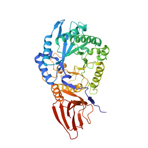
    A key component of colonization, biofilm formation, and protection of the opportunistic human pathogen Pseudomonas aeruginosa is the biosynthesis of the exopolysaccharide Psl. Composed of a pentameric repeating unit of mannose, glucose, and rhamnose, the biosynthesis of Psl is proposed to occur via a Wzx/Wzy-dependent mechanism. Previous genetic studies have shown that the putative glycoside hydrolase PslG is essential for Psl biosynthesis. To understand the function of this protein, the apo-structure of the periplasmic domain of PslG (PslG(31-442)) and its complex with mannose were determined to 2.0 and 1.9 Å resolution, respectively. Despite a domain architecture and positioning of catalytic residues similar to those of other family 39 glycoside hydrolases, PslG(31-442) exhibits a unique 32-Å-long active site groove that is distinct from other structurally characterized family members. PslG formed a complex with two mannose monosaccharides in this groove, consistent with binding data obtained from intrinsic tryptophan fluorescence. PslG was able to catalyze the hydrolysis of surface-associated Psl, and this activity was abolished in a E165Q/E276Q double catalytic variant. Surprisingly, P. aeruginosa variants with these chromosomal mutations as well as a pslG deletion mutant were still capable of forming Psl biofilms. However, overexpression of PslG in a pslG deletion background impaired biofilm formation and resulted in less surface-associated Psl, suggesting that regulation of this enzyme is important during polysaccharide biosynthesis.


  • Organizational Affiliation

    Program in Molecular Structure and Function, Research Institute, The Hospital for Sick Children, Toronto, Ontario M5G 1X8, Canada.


Macromolecules
Find similar proteins by:  (by identity cutoff)  |  3D Structure
Entity ID: 1
MoleculeChains Sequence LengthOrganismDetailsImage
PslG416Pseudomonas aeruginosa PAO1Mutation(s): 0 
Gene Names: pslGPA2237
UniProt
Find proteins for Q9I1N2 (Pseudomonas aeruginosa (strain ATCC 15692 / DSM 22644 / CIP 104116 / JCM 14847 / LMG 12228 / 1C / PRS 101 / PAO1))
Explore Q9I1N2 
Go to UniProtKB:  Q9I1N2
Entity Groups  
Sequence Clusters30% Identity50% Identity70% Identity90% Identity95% Identity100% Identity
UniProt GroupQ9I1N2
Sequence Annotations
Expand
  • Reference Sequence
Small Molecules
Ligands 4 Unique
IDChains Name / Formula / InChI Key2D Diagram3D Interactions
MAN
Query on MAN

Download Ideal Coordinates CCD File 
F [auth A],
G [auth A],
H [auth A],
I [auth A]
alpha-D-mannopyranose
C6 H12 O6
WQZGKKKJIJFFOK-PQMKYFCFSA-N
CD
Query on CD

Download Ideal Coordinates CCD File 
B [auth A],
C [auth A],
D [auth A],
E [auth A]
CADMIUM ION
Cd
WLZRMCYVCSSEQC-UHFFFAOYSA-N
EDO
Query on EDO

Download Ideal Coordinates CCD File 
K [auth A]1,2-ETHANEDIOL
C2 H6 O2
LYCAIKOWRPUZTN-UHFFFAOYSA-N
CL
Query on CL

Download Ideal Coordinates CCD File 
J [auth A]CHLORIDE ION
Cl
VEXZGXHMUGYJMC-UHFFFAOYSA-M
Experimental Data & Validation

Experimental Data

  • Method: X-RAY DIFFRACTION
  • Resolution: 1.90 Å
  • R-Value Free: 0.187 
  • R-Value Work: 0.155 
  • R-Value Observed: 0.156 
  • Space Group: P 41 21 2
Unit Cell:
Length ( Å )Angle ( ˚ )
a = 83.655α = 90
b = 83.655β = 90
c = 163.366γ = 90
Software Package:
Software NamePurpose
PHENIXrefinement
HKL-2000data reduction
HKL-2000data scaling
Cootmodel building
PHASERphasing

Structure Validation

View Full Validation Report


Ligand Structure Quality Assessment 


Entry History & Funding Information

Deposition Data


Funding OrganizationLocationGrant Number
Canadian Institutes of Health Research (CIHR)Canada43998

Revision History  (Full details and data files)

  • Version 1.0: 2015-10-07
    Type: Initial release
  • Version 1.1: 2015-10-14
    Changes: Database references
  • Version 1.2: 2015-12-02
    Changes: Database references
  • Version 1.3: 2017-11-01
    Changes: Author supporting evidence, Database references, Derived calculations
  • Version 1.4: 2020-01-08
    Changes: Author supporting evidence
  • Version 1.5: 2020-07-29
    Type: Remediation
    Reason: Carbohydrate remediation
    Changes: Data collection, Derived calculations, Structure summary
  • Version 1.6: 2023-09-27
    Changes: Data collection, Database references, Refinement description, Structure summary