5BWO

Crystal Structure of Human SIRT3 in Complex with a Palmitoyl H3K9 Peptide


Experimental Data Snapshot

  • Method: X-RAY DIFFRACTION
  • Resolution: 2.38 Å
  • R-Value Free: 0.287 
  • R-Value Work: 0.239 
  • R-Value Observed: 0.244 

Starting Model: experimental
View more details

wwPDB Validation   3D Report Full Report


Ligand Structure Quality Assessment 


This is version 1.2 of the entry. See complete history


Literature

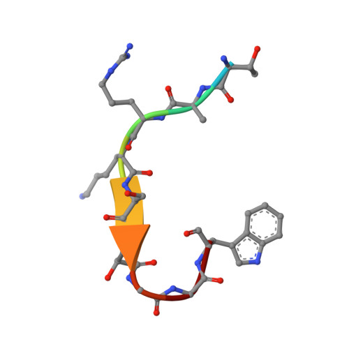
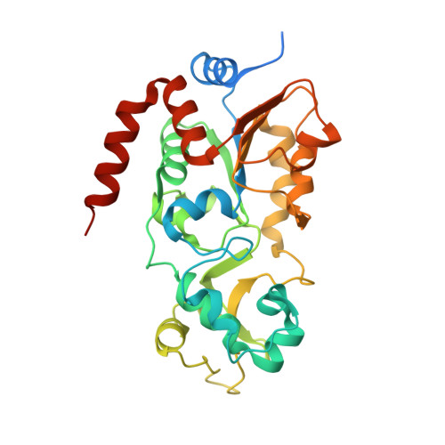
Crystal structures of SIRT3 reveal that the alpha 2-alpha 3 loop and alpha 3-helix affect the interaction with long-chain acyl lysine.

Gai, W.Li, H.Jiang, H.Long, Y.Liu, D.

(2016) FEBS Lett 590: 3019-3028

  • DOI: https://doi.org/10.1002/1873-3468.12345
  • Primary Citation of Related Structures:  
    5BWN, 5BWO

  • PubMed Abstract: 

    SIRT1-7 play important roles in many biological processes and age-related diseases. In addition to a NAD(+) -dependent deacetylase activity, they can catalyze several other reactions, including the hydrolysis of long-chain fatty acyl lysine. To study the binding modes of sirtuins to long-chain acyl lysines, we solved the crystal structures of SIRT3 bound to either a H3K9-myristoylated- or a H3K9-palmitoylated peptide. Interaction of SIRT3 with the palmitoyl group led to unfolding of the α3-helix. The myristoyl and palmitoyl groups bind to the C-pocket and an allosteric site near the α3-helix, respectively. We found that the residues preceding the α3-helix determine the size of the C-pocket. The flexibility of the α2-α3 loop and the plasticity of the α3-helix affect the interaction with long-chain acyl lysine.


  • Organizational Affiliation

    Department of Pharmacology III, Key Laboratory of Receptor Research, Shanghai Institute of Materia Medica, Chinese Academy of Sciences, China.


Macromolecules

Find similar proteins by:  Sequence   |   3D Structure  

Entity ID: 1
MoleculeChains Sequence LengthOrganismDetailsImage
Palmitoyl H3K9 PeptideA [auth B]10synthetic constructMutation(s): 0 
UniProt & NIH Common Fund Data Resources
Find proteins for Q6NXT2 (Homo sapiens)
Explore Q6NXT2 
Go to UniProtKB:  Q6NXT2
PHAROS:  Q6NXT2
GTEx:  ENSG00000188375 
Entity Groups  
Sequence Clusters30% Identity50% Identity70% Identity90% Identity95% Identity100% Identity
UniProt GroupQ6NXT2
Sequence Annotations
Expand
  • Reference Sequence
Find similar proteins by:  (by identity cutoff)  |  3D Structure
Entity ID: 2
MoleculeChains Sequence LengthOrganismDetailsImage
NAD-dependent protein deacetylase sirtuin-3, mitochondrialB [auth A]309Homo sapiensMutation(s): 0 
Gene Names: SIRT3SIR2L3
EC: 3.5.1
UniProt & NIH Common Fund Data Resources
Find proteins for Q9NTG7 (Homo sapiens)
Explore Q9NTG7 
Go to UniProtKB:  Q9NTG7
GTEx:  ENSG00000142082 
Entity Groups  
Sequence Clusters30% Identity50% Identity70% Identity90% Identity95% Identity100% Identity
UniProt GroupQ9NTG7
Sequence Annotations
Expand
  • Reference Sequence
Small Molecules
Experimental Data & Validation

Experimental Data

  • Method: X-RAY DIFFRACTION
  • Resolution: 2.38 Å
  • R-Value Free: 0.287 
  • R-Value Work: 0.239 
  • R-Value Observed: 0.244 
  • Space Group: P 21 21 21
Unit Cell:
Length ( Å )Angle ( ˚ )
a = 43.445α = 90
b = 54.636β = 90
c = 113.203γ = 90
Software Package:
Software NamePurpose
PHENIXrefinement
HKL-2000data scaling
PHENIXmodel building

Structure Validation

View Full Validation Report


Ligand Structure Quality Assessment 


Entry History 

Deposition Data

Revision History  (Full details and data files)

  • Version 1.0: 2016-07-13
    Type: Initial release
  • Version 1.1: 2016-12-07
    Changes: Database references
  • Version 1.2: 2023-11-08
    Changes: Data collection, Database references, Refinement description