5BR7

Structure of UDP-galactopyranose mutase from Corynebacterium diphtheriae in complex with citrate ion


Experimental Data Snapshot

  • Method: X-RAY DIFFRACTION
  • Resolution: 1.95 Å
  • R-Value Free: 0.190 
  • R-Value Work: 0.153 
  • R-Value Observed: 0.155 

wwPDB Validation   3D Report Full Report


Ligand Structure Quality Assessment 


This is version 1.2 of the entry. See complete history


Literature

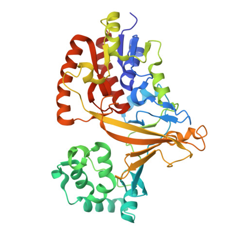
Conformational Control of UDP-Galactopyranose Mutase Inhibition.

Wangkanont, K.Winton, V.J.Forest, K.T.Kiessling, L.L.

(2017) Biochemistry 56: 3983-3992

  • DOI: https://doi.org/10.1021/acs.biochem.7b00189
  • Primary Citation of Related Structures:  
    5BR7, 5EQF

  • PubMed Abstract: 

    UDP-galactopyranose mutase (Glf or UGM) catalyzes the formation of uridine 5'-diphosphate-α-d-galactofuranose (UDP-Galf) from UDP-galactopyranose (UDP-Galp). The enzyme is required for the production of Galf-containing glycans. UGM is absent in mammals, but members of the Corynebacterineae suborder require UGM for cell envelope biosynthesis. The need for UGM in some pathogens has prompted the search for inhibitors that could serve as antibiotic leads. Optimizing inhibitor potency, however, has been challenging. The UGM from Klebsiella pneumoniae (KpUGM), which is not required for viability, is more effectively impeded by small-molecule inhibitors than are essential UGMs from species such as Mycobacterium tuberculosis or Corynebacterium diphtheriae. Why KpUGM is more susceptible to inhibition than other orthologs is not clear. One potential source of difference is UGM ortholog conformation. We previously determined a structure of CdUGM bound to a triazolothiadiazine inhibitor in the open form, but it was unclear whether the small-molecule inhibitor bound this form or to the closed form. By varying the terminal tag (CdUGM-His 6 and GSG-CdUGM), we crystallized CdUGM to capture the enzyme in different conformations. These structures reveal a pocket in the active site that can be exploited to augment inhibitor affinity. Moreover, they suggest the inhibitor binds the open form of most prokaryotic UGMs but can bind the closed form of KpUGM. This model and the structures suggest strategies for optimizing inhibitor potency by exploiting UGM conformational flexibility.


  • Organizational Affiliation

    Department of Chemistry, ‡Department of Biochemistry, and §Department of Bacteriology, University of Wisconsin-Madison , Madison, Wisconsin 53706, United States.


Macromolecules
Find similar proteins by:  (by identity cutoff)  |  3D Structure
Entity ID: 1
MoleculeChains Sequence LengthOrganismDetailsImage
UDP-galactopyranose mutase393Corynebacterium diphtheriae NCTC 13129Mutation(s): 0 
Gene Names: glfDIP2203
EC: 5.4.99.9
UniProt
Find proteins for Q6NER4 (Corynebacterium diphtheriae (strain ATCC 700971 / NCTC 13129 / Biotype gravis))
Explore Q6NER4 
Go to UniProtKB:  Q6NER4
Entity Groups  
Sequence Clusters30% Identity50% Identity70% Identity90% Identity95% Identity100% Identity
UniProt GroupQ6NER4
Sequence Annotations
Expand
  • Reference Sequence
Small Molecules
Ligands 5 Unique
IDChains Name / Formula / InChI Key2D Diagram3D Interactions
FAD
Query on FAD

Download Ideal Coordinates CCD File 
B [auth A]FLAVIN-ADENINE DINUCLEOTIDE
C27 H33 N9 O15 P2
VWWQXMAJTJZDQX-UYBVJOGSSA-N
FLC
Query on FLC

Download Ideal Coordinates CCD File 
L [auth A]CITRATE ANION
C6 H5 O7
KRKNYBCHXYNGOX-UHFFFAOYSA-K
EDO
Query on EDO

Download Ideal Coordinates CCD File 
C [auth A],
D [auth A],
E [auth A],
F [auth A]
1,2-ETHANEDIOL
C2 H6 O2
LYCAIKOWRPUZTN-UHFFFAOYSA-N
IPA
Query on IPA

Download Ideal Coordinates CCD File 
I [auth A],
J [auth A],
K [auth A]
ISOPROPYL ALCOHOL
C3 H8 O
KFZMGEQAYNKOFK-UHFFFAOYSA-N
NA
Query on NA

Download Ideal Coordinates CCD File 
G [auth A],
H [auth A]
SODIUM ION
Na
FKNQFGJONOIPTF-UHFFFAOYSA-N
Experimental Data & Validation

Experimental Data

  • Method: X-RAY DIFFRACTION
  • Resolution: 1.95 Å
  • R-Value Free: 0.190 
  • R-Value Work: 0.153 
  • R-Value Observed: 0.155 
  • Space Group: P 41 21 2
Unit Cell:
Length ( Å )Angle ( ˚ )
a = 98.278α = 90
b = 98.278β = 90
c = 126.223γ = 90
Software Package:
Software NamePurpose
SCALEPACKdata scaling
PHASERphasing
PHENIXrefinement
PDB_EXTRACTdata extraction
HKL-2000data reduction

Structure Validation

View Full Validation Report


Ligand Structure Quality Assessment 


Entry History 

Deposition Data

Revision History  (Full details and data files)

  • Version 1.0: 2016-06-01
    Type: Initial release
  • Version 1.1: 2017-08-09
    Changes: Database references, Derived calculations
  • Version 1.2: 2023-09-27
    Changes: Data collection, Database references, Refinement description