Structural basis of the interaction between the meningitis pathogen Streptococcus suis adhesin Fhb and its human receptor.
Zhang, C., Hao, H., Yu, Y., Kong, D., Chen, S., Jiang, H., Yuan, Y., Zheng, Y., Yang, M., Jiang, Y.(2016) FEBS Lett 590: 1384-1392
- PubMed: 27086582 Search on PubMed
- DOI: https://doi.org/10.1002/1873-3468.12174
- Primary Citation Related Structures:
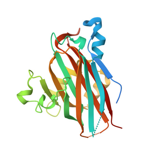
5BOA - PubMed Abstract:
The recently identified Streptococcus suis adhesin factor H-binding protein (Fhb) targets the host cellular receptor glycolipid GbO3 through its N terminus. However, it is unclear how Fhb interacts with its receptor. Here, we determined the complex structure of factor H-binding protein receptor-binding domain (Fhb RBD) with Gb2, an analog of its receptor, revealing that Gb2 binds in a pocket of the β sandwich core domain. We identified the key residues for Fhb RBD receptor binding using mutagenesis and isothermal titration calorimetry. Mutagenesis analyses indicated that Fhb binds to Gb2 mainly through hydrogen and hydrophobic interactions. Our findings provided structural insights into the Fhb-mediated host-pathogen interactions of S. suis.
- State Key Laboratory of Pathogen and Biosecurity, Beijing Institute of Microbiology and Epidemiology, China.
Organizational Affiliation: