5BJP

Crystal structure of the Corn RNA aptamer in complex with DFHO, iridium hexammine soak


Experimental Data Snapshot

  • Method: X-RAY DIFFRACTION
  • Resolution: 2.51 Å
  • R-Value Free: 0.239 
  • R-Value Work: 0.191 
  • R-Value Observed: 0.195 

wwPDB Validation   3D Report Full Report


Ligand Structure Quality Assessment 


This is version 1.3 of the entry. See complete history


Literature

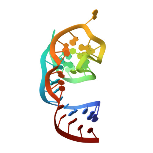
A homodimer interface without base pairs in an RNA mimic of red fluorescent protein.

Warner, K.D.Sjekloca, L.Song, W.Filonov, G.S.Jaffrey, S.R.Ferre-D'Amare, A.R.

(2017) Nat Chem Biol 13: 1195-1201

  • DOI: https://doi.org/10.1038/nchembio.2475
  • Primary Citation of Related Structures:  
    5BJO, 5BJP

  • PubMed Abstract: 

    Corn, a 28-nucleotide RNA, increases yellow fluorescence of its cognate ligand 3,5-difluoro-4-hydroxybenzylidene-imidazolinone-2-oxime (DFHO) by >400-fold. Corn was selected in vitro to overcome limitations of other fluorogenic RNAs, particularly rapid photobleaching. We now report the Corn-DFHO co-crystal structure, discovering that the functional species is a quasisymmetric homodimer. Unusually, the dimer interface, in which six unpaired adenosines break overall two-fold symmetry, lacks any intermolecular base pairs. The homodimer encapsulates one DFHO at its interprotomer interface, sandwiching it with a G-quadruplex from each protomer. Corn and the green-fluorescent Spinach RNA are structurally unrelated. Their convergent use of G-quadruplexes underscores the usefulness of this motif for RNA-induced small-molecule fluorescence. The asymmetric dimer interface of Corn could provide a basis for the development of mutants that only fluoresce as heterodimers. Such variants would be analogous to Split GFP, and may be useful for analyzing RNA co-expression or association, or for designing self-assembling RNA nanostructures.


  • Organizational Affiliation

    Biochemistry and Biophysics Center, National Heart, Lung and Blood Institute, Bethesda, Maryland, USA.


Macromolecules
Find similar nucleic acids by:  (by identity cutoff)  |  3D Structure
Entity ID: 1
MoleculeChains LengthOrganismImage
RNA (36-MER)A [auth E],
B [auth Y]
36synthetic construct
Sequence Annotations
Expand
  • Reference Sequence
Small Molecules
Ligands 5 Unique
IDChains Name / Formula / InChI Key2D Diagram3D Interactions
747
Query on 747

Download Ideal Coordinates CCD File 
L [auth Y](5Z)-5-[(3,5-difluoro-4-hydroxyphenyl)methylidene]-2-[(E)-(hydroxyimino)methyl]-3-methyl-3,5-dihydro-4H-imidazol-4-one
C12 H9 F2 N3 O3
MZFHSQVSFUWKDU-TUUOAFJKSA-N
IR
Query on IR

Download Ideal Coordinates CCD File 
C [auth E],
I [auth Y]
IRIDIUM ION
Ir
MOHYGSBMXIJZBJ-UHFFFAOYSA-N
DMS
Query on DMS

Download Ideal Coordinates CCD File 
H [auth E]DIMETHYL SULFOXIDE
C2 H6 O S
IAZDPXIOMUYVGZ-UHFFFAOYSA-N
K
Query on K

Download Ideal Coordinates CCD File 
D [auth E],
E,
J [auth Y],
K [auth Y]
POTASSIUM ION
K
NPYPAHLBTDXSSS-UHFFFAOYSA-N
MG
Query on MG

Download Ideal Coordinates CCD File 
F [auth E],
G [auth E]
MAGNESIUM ION
Mg
JLVVSXFLKOJNIY-UHFFFAOYSA-N
Experimental Data & Validation

Experimental Data

  • Method: X-RAY DIFFRACTION
  • Resolution: 2.51 Å
  • R-Value Free: 0.239 
  • R-Value Work: 0.191 
  • R-Value Observed: 0.195 
  • Space Group: P 21 21 21
Unit Cell:
Length ( Å )Angle ( ˚ )
a = 36.642α = 90
b = 55.438β = 90
c = 98.676γ = 90
Software Package:
Software NamePurpose
REFMACrefinement
HKL-2000data reduction
HKL-2000data scaling
AutoSolphasing

Structure Validation

View Full Validation Report


Ligand Structure Quality Assessment 


Entry History 

Deposition Data

Revision History  (Full details and data files)

  • Version 1.0: 2017-09-27
    Type: Initial release
  • Version 1.1: 2017-10-11
    Changes: Database references
  • Version 1.2: 2017-10-25
    Changes: Database references
  • Version 1.3: 2024-03-06
    Changes: Data collection, Database references, Derived calculations