


Crystal Structure of Type III pantothenate kinase (PanK III) from Burkholderia cenocepacia complexed with pantothenate, imidodiphosphate, and AMP
SSGCID, Dranow, D.M., Abendroth, J., Lorimer, D., Edwards, T.E.To be published.
Experimental Data Snapshot
Starting Model: experimental
View more details
Entity ID: 1 | |||||
|---|---|---|---|---|---|
| Molecule | Chains | Sequence Length | Organism | Details | Image |
| Type III pantothenate kinase | 290 | Burkholderia cenocepacia J2315 | Mutation(s): 0 Gene Names: coaX, BceJ2315_06870, BCAL0693 EC: 2.7.1.33 | 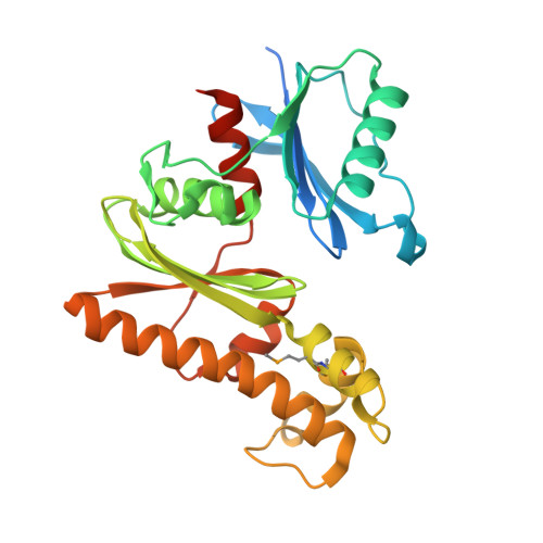 | |
UniProt | |||||
Entity Groups | |||||
| Sequence Clusters | 30% Identity50% Identity70% Identity90% Identity95% Identity100% Identity | ||||
| UniProt Group | B4E9P3 | ||||
Sequence AnnotationsExpand | |||||
Reference Sequence | |||||
| Ligands 4 Unique | |||||
|---|---|---|---|---|---|
| ID | Chains | Name / Formula / InChI Key | 2D Diagram | 3D Interactions | |
| AMP Download:Ideal Coordinates CCD File | F [auth A] | ADENOSINE MONOPHOSPHATE C10 H14 N5 O7 P UDMBCSSLTHHNCD-KQYNXXCUSA-N |  | ||
| PAU Download:Ideal Coordinates CCD File | D [auth A], H [auth B] | PANTOTHENOIC ACID C9 H17 N O5 GHOKWGTUZJEAQD-ZETCQYMHSA-N |  | ||
| 2PN Download:Ideal Coordinates CCD File | C [auth A], G [auth B] | IMIDODIPHOSPHORIC ACID H5 N O6 P2 GNGSOPFGGKKDQP-UHFFFAOYSA-N |  | ||
| EDO Download:Ideal Coordinates CCD File | E [auth A], I [auth B] | 1,2-ETHANEDIOL C2 H6 O2 LYCAIKOWRPUZTN-UHFFFAOYSA-N |  | ||
| Modified Residues 1 Unique | |||||
|---|---|---|---|---|---|
| ID | Chains | Type | Formula | 2D Diagram | Parent |
| MSE Query on MSE | A, B | L-PEPTIDE LINKING | C5 H11 N O2 Se |  | MET |
| Length ( Å ) | Angle ( ˚ ) |
|---|---|
| a = 44.21 | α = 90 |
| b = 72.75 | β = 90 |
| c = 154.02 | γ = 90 |
| Software Name | Purpose |
|---|---|
| XSCALE | data scaling |
| PHASER | phasing |
| PHENIX | refinement |
| PDB_EXTRACT | data extraction |
| XDS | data reduction |
| ARP | model building |


