5B6M | pdb_00005b6m

Crystal structure of human peroxiredoxin 6 in reduced state


Experimental Data Snapshot

  • Method: X-RAY DIFFRACTION
  • Resolution: 2.50 Å
  • R-Value Free: 
    0.290 (Depositor), 0.238 (DCC) 
  • R-Value Work: 
    0.233 (Depositor), 0.237 (DCC) 
  • R-Value Observed: 
    0.235 (Depositor) 

Starting Model: experimental
View more details

wwPDB Validation 3D Report Full Report

Validation slider image for 5B6M

This is version 1.2 of the entry. See complete history

Literature

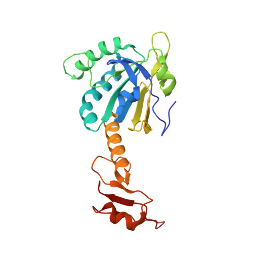
Crystal structures of human peroxiredoxin 6 in different oxidation states

Kim, K.H.Lee, W.T.Kim, E.E.

(2016) Biochem Biophys Res Commun 477: 717-722

  • DOI: https://doi.org/10.1016/j.bbrc.2016.06.125
  • Primary Citation Related Structures: 
    5B6M, 5B6N

  • PubMed Abstract: 

    Peroxiredoxins (Prxs) are a family of antioxidant enzymes found ubiquitously. Prxs function not only as H2O2 scavengers but also as highly sensitive H2O2 sensors and signal transducers. Since reactive oxygen species are involved in many cellular metabolic and signaling processes, Prxs play important roles in various diseases. Prxs can be hyperoxidized to the sulfinic acid (SO2H) or sulfonic acid (SO3H) forms in the presence of high concentrations of H2O2. It is known that oligomerization of Prx is changed accompanying oxidation states, and linked to the function. Among the six Prxs in mammals, Prx6 is the only 1-Cys Prx. It is found in all organs in humans, unlike some 2-Cys Prxs, and is present in all species from bacteria to humans. In addition, Prx6 has Ca(2+)-independent phospholipase A2 (PLA2) activity. Thus far only the crystal structure of Prx in the oxidized state has been reported. In this study, we present the crystal structures of human Prx6 in the reduced (SH) and the sulfinic acid (SO2H) forms.


  • Organizational Affiliation
    • Biomedical Research Institute, Korea Institute of Science and Technology, Seoul 02792, Republic of Korea; Department of Biochemistry, College of Life Science and Biotechnology, Yonsei University, Seoul 03722, Republic of Korea.

Macromolecule Content 

  • Total Structure Weight: 150.41 kDa 
  • Atom Count: 10,485 
  • Modeled Residue Count: 1,311 
  • Deposited Residue Count: 1,344 
  • Unique protein chains: 1

Macromolecules

Find similar proteins by:|  3D Structure
Entity ID: 1
MoleculeChains  Sequence LengthOrganismDetailsImage
Peroxiredoxin-6
A, B, C, D, E
A, B, C, D, E, F
224Homo sapiensMutation(s): 0 
Gene Names: PRDX6AOP2KIAA0106
EC: 1.11.1.15 (PDB Primary Data), 3.1.1.4 (UniProt), 2.3.1.23 (UniProt), 1.11.1.27 (UniProt)
UniProt & NIH Common Fund Data Resources
Find proteins for P30041 (Homo sapiens)
Explore P30041 
Go to UniProtKB:  P30041
PHAROS:  P30041
GTEx:  ENSG00000117592 
Entity Groups
Sequence Clusters30% Identity50% Identity70% Identity90% Identity95% Identity100% Identity
UniProt GroupP30041
Sequence Annotations
Expand
Reference Sequence

Experimental Data & Validation

Experimental Data

  • Method: X-RAY DIFFRACTION
  • Resolution: 2.50 Å
  • R-Value Free:  0.290 (Depositor), 0.238 (DCC) 
  • R-Value Work:  0.233 (Depositor), 0.237 (DCC) 
  • R-Value Observed: 0.235 (Depositor) 
Space Group: P 21 21 21
Unit Cell:
Length ( Å )Angle ( ˚ )
a = 94.353α = 90
b = 106.349β = 90
c = 165.504γ = 90
Software Package:
Software NamePurpose
PHENIXrefinement
HKL-2000data reduction
HKL-2000data scaling
CNSphasing

Structure Validation

View Full Validation Report


Entry History 

& Funding Information

Deposition Data


Funding OrganizationLocationGrant Number
Ministry of Science, ICT and Future Planning of KoreaKorea, Republic OfNRF 20110021713

Revision History  (Full details and data files)

  • Version 1.0: 2016-07-13
    Type: Initial release
  • Version 1.1: 2016-08-10
    Changes: Database references
  • Version 1.2: 2023-11-08
    Changes: Data collection, Database references, Derived calculations, Refinement description