5B4W

Crystal structure of Plexin inhibitor complex


Experimental Data Snapshot

  • Method: X-RAY DIFFRACTION
  • Resolution: 2.60 Å
  • R-Value Free: 0.265 
  • R-Value Work: 0.225 
  • R-Value Observed: 0.227 

wwPDB Validation   3D Report Full Report


Ligand Structure Quality Assessment 


This is version 1.4 of the entry. See complete history


Literature

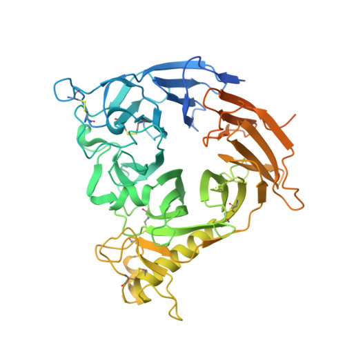
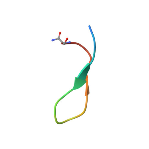
Allosteric Inhibition of a Semaphorin 4D Receptor Plexin B1 by a High-Affinity Macrocyclic Peptide

Matsunaga, Y.Bashiruddin, N.K.Kitago, Y.Takagi, J.Suga, H.

(2016) Cell Chem Biol 23: 1341-1350

  • DOI: https://doi.org/10.1016/j.chembiol.2016.09.015
  • Primary Citation of Related Structures:  
    5B4W

  • PubMed Abstract: 

    Semaphorin axonal guidance factors are multifunctional proteins that play important roles in immune response, cancer cell proliferation, and organogenesis, making semaphorins and their signaling receptor plexins important drug targets for various diseases. However, the large and flat binding surface of the semaphorin-plexin interaction interface is difficult to target by traditional small-molecule drugs. Here, we report the discovery of a high-affinity plexin B1 (PlxnB1)-binding macrocyclic peptide, PB1m6 (K D  = 3.5 nM). PB1m6 specifically inhibited the binding of physiological ligand semaphorin 4D (Sema4D) in vitro and completely suppressed Sema4D-induced cell collapse. Structural studies revealed that PB1m6 binds at a groove between the fifth and sixth blades of the sema domain in PlxnB1 distant from the Sema4D-binding site, indicating the non-competitive and allosteric nature of the inhibitory activity. The discovery of this novel allosteric site can potentially be used to target plexin family proteins for the development of drugs that modulate semaphorin and plexin signaling.


  • Organizational Affiliation

    Laboratory of Protein Synthesis and Expression, Institute for Protein Research, Osaka University, Osaka 565-0871, Japan.


Macromolecules
Find similar proteins by:  (by identity cutoff)  |  3D Structure
Entity ID: 1
MoleculeChains Sequence LengthOrganismDetailsImage
Plexin-B1
A, B, C, D, E
A, B, C, D, E, F
579Homo sapiensMutation(s): 0 
Gene Names: PLXNB1KIAA0407PLXN5SEP
UniProt & NIH Common Fund Data Resources
Find proteins for O43157 (Homo sapiens)
Explore O43157 
Go to UniProtKB:  O43157
PHAROS:  O43157
GTEx:  ENSG00000164050 
Entity Groups  
Sequence Clusters30% Identity50% Identity70% Identity90% Identity95% Identity100% Identity
UniProt GroupO43157
Sequence Annotations
Expand
  • Reference Sequence

Find similar proteins by:  Sequence   |   3D Structure  

Entity ID: 2
MoleculeChains Sequence LengthOrganismDetailsImage
Synthesized cyclic peptide
G, H, I, J, K
G, H, I, J, K, L
18unidentifiedMutation(s): 0 
Entity Groups  
Sequence Clusters30% Identity50% Identity70% Identity90% Identity95% Identity100% Identity
Sequence Annotations
Expand
  • Reference Sequence
Experimental Data & Validation

Experimental Data

  • Method: X-RAY DIFFRACTION
  • Resolution: 2.60 Å
  • R-Value Free: 0.265 
  • R-Value Work: 0.225 
  • R-Value Observed: 0.227 
  • Space Group: P 1 21 1
Unit Cell:
Length ( Å )Angle ( ˚ )
a = 92.426α = 90
b = 217.924β = 115.3
c = 106.475γ = 90
Software Package:
Software NamePurpose
PHENIXrefinement
DENZOdata collection
SCALEPACKdata scaling
PHASERphasing

Structure Validation

View Full Validation Report


Ligand Structure Quality Assessment 


Entry History & Funding Information

Deposition Data


Funding OrganizationLocationGrant Number
AMEDJapan--

Revision History  (Full details and data files)

  • Version 1.0: 2016-12-28
    Type: Initial release
  • Version 1.1: 2020-02-26
    Changes: Data collection
  • Version 1.2: 2020-07-29
    Type: Remediation
    Reason: Carbohydrate remediation
    Changes: Data collection, Derived calculations, Structure summary
  • Version 1.3: 2023-11-08
    Changes: Data collection, Database references, Refinement description, Structure summary
  • Version 1.4: 2023-11-15
    Changes: Data collection