5B3U

Crystal structure of biliverdin reductase in complex with NADP+ from Synechocystis sp. PCC 6803


Experimental Data Snapshot

  • Method: X-RAY DIFFRACTION
  • Resolution: 2.70 Å
  • R-Value Free: 0.271 
  • R-Value Work: 0.223 
  • R-Value Observed: 0.225 

wwPDB Validation   3D Report Full Report


Ligand Structure Quality Assessment 


This is version 1.4 of the entry. See complete history


Literature

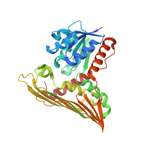
A substrate-bound structure of cyanobacterial biliverdin reductase identifies stacked substrates as critical for activity

Takao, H.Hirabayashi, K.Nishigaya, Y.Kouriki, H.Nakaniwa, T.Hagiwara, Y.Harada, J.Sato, H.Yamazaki, T.Sakakibara, Y.Suiko, M.Asada, Y.Takahashi, Y.Yamamoto, K.Fukuyama, K.Sugishima, M.Wada, K.

(2017) Nat Commun 8: 14397-14397

  • DOI: https://doi.org/10.1038/ncomms14397
  • Primary Citation of Related Structures:  
    5B3T, 5B3U, 5B3V

  • PubMed Abstract: 

    Biliverdin reductase catalyses the last step in haem degradation and produces the major lipophilic antioxidant bilirubin via reduction of biliverdin, using NAD(P)H as a cofactor. Despite the importance of biliverdin reductase in maintaining the redox balance, the molecular details of the reaction it catalyses remain unknown. Here we present the crystal structure of biliverdin reductase in complex with biliverdin and NADP + . Unexpectedly, two biliverdin molecules, which we designated the proximal and distal biliverdins, bind with stacked geometry in the active site. The nicotinamide ring of the NADP + is located close to the reaction site on the proximal biliverdin, supporting that the hydride directly attacks this position of the proximal biliverdin. The results of mutagenesis studies suggest that a conserved Arg185 is essential for the catalysis. The distal biliverdin probably acts as a conduit to deliver the proton from Arg185 to the proximal biliverdin, thus yielding bilirubin.


  • Organizational Affiliation

    Organization for Promotion of Tenure Track, University of Miyazaki, Miyazaki 889-1692, Japan.


Macromolecules
Find similar proteins by:  (by identity cutoff)  |  3D Structure
Entity ID: 1
MoleculeChains Sequence LengthOrganismDetailsImage
Biliverdin reductase
A, B
331Synechocystis sp. PCC 6803 substr. KazusaMutation(s): 0 
Gene Names: bvdR
UniProt
Find proteins for P72782 (Synechocystis sp. (strain PCC 6803 / Kazusa))
Explore P72782 
Go to UniProtKB:  P72782
Entity Groups  
Sequence Clusters30% Identity50% Identity70% Identity90% Identity95% Identity100% Identity
UniProt GroupP72782
Sequence Annotations
Expand
  • Reference Sequence
Experimental Data & Validation

Experimental Data

  • Method: X-RAY DIFFRACTION
  • Resolution: 2.70 Å
  • R-Value Free: 0.271 
  • R-Value Work: 0.223 
  • R-Value Observed: 0.225 
  • Space Group: P 21 21 21
Unit Cell:
Length ( Å )Angle ( ˚ )
a = 59.091α = 90
b = 85.562β = 90
c = 135.026γ = 90
Software Package:
Software NamePurpose
PHENIXrefinement
XDSdata scaling
PHENIXphasing
Cootmodel building
XDSdata reduction

Structure Validation

View Full Validation Report


Ligand Structure Quality Assessment 


Entry History 

Deposition Data

Revision History  (Full details and data files)

  • Version 1.0: 2017-02-15
    Type: Initial release
  • Version 1.1: 2017-02-22
    Changes: Database references
  • Version 1.2: 2020-02-26
    Changes: Data collection
  • Version 1.3: 2021-08-11
    Changes: Database references, Structure summary
  • Version 1.4: 2023-11-08
    Changes: Data collection, Refinement description