


Crystal Structure of P450BM3 with decoy molecules
Cong, Z., Shoji, O., Kasai, C., Sugimoto, H., Shiro, Y., Watanabe, Y.To be published.
Experimental Data Snapshot
Starting Model: experimental
View more details
Entity ID: 1 | |||||
|---|---|---|---|---|---|
| Molecule | Chains | Sequence Length | Organism | Details | Image |
| Bifunctional cytochrome P450/NADPH--P450 reductase | 456 | Priestia megaterium | Mutation(s): 1 Gene Names: cyp102A1, cyp102 EC: 1.14.14.1 (PDB Primary Data), 1.6.2.4 (PDB Primary Data) | 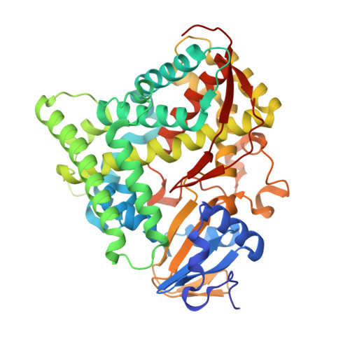 | |
UniProt | |||||
Entity Groups | |||||
| Sequence Clusters | 30% Identity50% Identity70% Identity90% Identity95% Identity100% Identity | ||||
| UniProt Group | P14779 | ||||
Sequence AnnotationsExpand | |||||
Reference Sequence | |||||
| Ligands 3 Unique | |||||
|---|---|---|---|---|---|
| ID | Chains | Name / Formula / InChI Key | 2D Diagram | 3D Interactions | |
| HEM Download:Ideal Coordinates CCD File | C [auth A], F [auth B] | PROTOPORPHYRIN IX CONTAINING FE C34 H32 Fe N4 O4 KABFMIBPWCXCRK-RGGAHWMASA-L |  | ||
| W06 Download:Ideal Coordinates CCD File | D [auth A], G [auth B] | (2~{S})-3-(1~{H}-indol-3-yl)-2-[2,2,3,3,4,4,5,5,6,6,6-undecakis(fluoranyl)hexanoylamino]propanoic acid C17 H11 F11 N2 O3 HOHTXGBLBJYLPP-JTQLQIEISA-N |  | ||
| DMS Download:Ideal Coordinates CCD File | E [auth A], H [auth B] | DIMETHYL SULFOXIDE C2 H6 O S IAZDPXIOMUYVGZ-UHFFFAOYSA-N |  | ||
| Length ( Å ) | Angle ( ˚ ) |
|---|---|
| a = 58.473 | α = 90 |
| b = 144.893 | β = 97.06 |
| c = 62.522 | γ = 90 |
| Software Name | Purpose |
|---|---|
| DENZO | data collection |
| SCALEPACK | data scaling |
| MOLREP | phasing |
| REFMAC | refinement |
| PDB_EXTRACT | data extraction |
| Funding Organization | Location | Grant Number |
|---|---|---|
| Japan | -- | |
| -- |


