5B1W

Crystal structure of human dendritic cell inhibitory receptor (DCIR) C-type lectin domain in ligand-free form


Experimental Data Snapshot

  • Method: X-RAY DIFFRACTION
  • Resolution: 3.05 Å
  • R-Value Free: 0.284 
  • R-Value Work: 0.253 
  • R-Value Observed: 0.254 

wwPDB Validation   3D Report Full Report


This is version 1.2 of the entry. See complete history


Literature

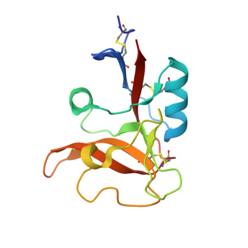
Crystal structure of human dendritic cell inhibitory receptor C-type lectin domain reveals the binding mode with N-glycan

Nagae, M.Ikeda, A.Hanashima, S.Kojima, T.Matsumoto, N.Yamamoto, K.Yamaguchi, Y.

(2016) FEBS Lett 590: 1280-1288

  • DOI: https://doi.org/10.1002/1873-3468.12162
  • Primary Citation of Related Structures:  
    5B1W, 5B1X

  • PubMed Abstract: 

    Human dendritic cell inhibitory receptor (DCIR) is a C-type lectin receptor expressed in classical dendritic cells and accepts several oligosaccharide ligands including N-glycans. Here, we report the crystal structures of human DCIR C-type lectin domains in the absence and presence of a branched N-glycan unit. The domain has a typical C-type lectin fold and two bound calcium ions. In the ligand-bound form, the disaccharide unit (GlcNAcβ1-2Man) acceptably fits the electron density map, indicating that it forms the main epitope. The recognition of the nonterminal N-glycan unit explains the relatively broad specificity of this lectin.


  • Organizational Affiliation

    Structural Glycobiology Team, Systems Glycobiology Research Group, RIKEN-Max Planck Joint Research Center, RIKEN Global Research Cluster, Wako, Saitama, Japan.


Macromolecules
Find similar proteins by:  (by identity cutoff)  |  3D Structure
Entity ID: 1
MoleculeChains Sequence LengthOrganismDetailsImage
C-type lectin domain family 4 member A
A, B, C, D
134Homo sapiensMutation(s): 0 
Gene Names: CLEC4ACLECSF6DCIRLLIRHDCGC13P
UniProt & NIH Common Fund Data Resources
Find proteins for Q9UMR7 (Homo sapiens)
Explore Q9UMR7 
Go to UniProtKB:  Q9UMR7
PHAROS:  Q9UMR7
GTEx:  ENSG00000111729 
Entity Groups  
Sequence Clusters30% Identity50% Identity70% Identity90% Identity95% Identity100% Identity
UniProt GroupQ9UMR7
Sequence Annotations
Expand
  • Reference Sequence
Experimental Data & Validation

Experimental Data

  • Method: X-RAY DIFFRACTION
  • Resolution: 3.05 Å
  • R-Value Free: 0.284 
  • R-Value Work: 0.253 
  • R-Value Observed: 0.254 
  • Space Group: P 21 21 2
Unit Cell:
Length ( Å )Angle ( ˚ )
a = 102.489α = 90
b = 105.832β = 90
c = 65.387γ = 90
Software Package:
Software NamePurpose
PHENIXrefinement
HKL-2000data reduction
HKL-2000data scaling
MOLREPphasing

Structure Validation

View Full Validation Report


Entry History & Funding Information

Deposition Data


Funding OrganizationLocationGrant Number
MEXTJapan24770111
MEXTJapan15K18496

Revision History  (Full details and data files)

  • Version 1.0: 2016-05-11
    Type: Initial release
  • Version 1.1: 2020-02-26
    Changes: Data collection, Database references, Derived calculations
  • Version 1.2: 2023-11-08
    Changes: Data collection, Database references, Derived calculations, Refinement description