5AY6 | pdb_00005ay6

32 kDa Fragment of the Flagellar hook protein FlgE from Caulobacter crescentus


Experimental Data Snapshot

  • Method: X-RAY DIFFRACTION
  • Resolution: 1.84 Å
  • R-Value Free: 
    0.222 (Depositor), 0.219 (DCC) 
  • R-Value Work: 
    0.176 (Depositor), 0.177 (DCC) 
  • R-Value Observed: 
    0.178 (Depositor) 

wwPDB Validation 3D Report Full Report

Validation slider image for 5AY6

This is version 1.4 of the entry. See complete history

Literature

Structural insights into bacterial flagellar hooks similarities and specificities

Yoon, Y.-H.Barker, C.S.Bulieris, P.V.Matsunami, H.Samatey, F.A.

(2016) Sci Rep 6: 35552-35552

  • DOI: https://doi.org/10.1038/srep35552
  • Primary Citation Related Structures: 
    5AY6, 5AZ4

  • PubMed Abstract: 

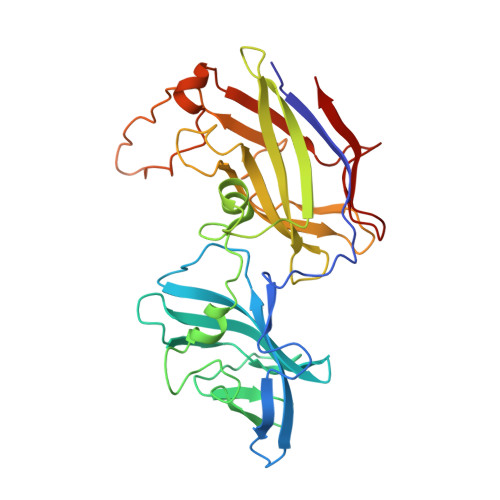
    Across bacteria, the protein that makes the flagellar hook, FlgE, has a high variability in amino acid residue composition and sequence length. We hereby present the structure of two fragments of FlgE protein from Campylobacter jejuni and from Caulobacter crescentus, which were obtained by X-ray crystallography, and a high-resolution model of the hook from Caulobacter. By comparing these new structures of FlgE proteins, we show that bacterial hook can be divided in two distinct parts. The first part comprises domains that are found in all FlgE proteins and that will make the basic structure of the hook that is common to all flagellated bacteria. The second part, hyper-variable both in size and structure, will be bacteria dependent. To have a better understanding of the C. jejuni hook, we show that a special strain of Salmonella enterica, which was designed to encode a gene of flgE that has the extra domains found in FlgE from C. jejuni, is fully motile. It seems that no matter the size of the hook protein, the hook will always have a structure made of 11 protofilaments.


  • Organizational Affiliation
    • Trans-Membrane Trafficking Unit, Okinawa Institute of Science and Technology Graduate University, 1919-1, Tancha, Onna, Kunigami, Okinawa 904-0495, Japan.

Macromolecule Content 

  • Total Structure Weight: 63.15 kDa 
  • Atom Count: 5,516 
  • Modeled Residue Count: 606 
  • Deposited Residue Count: 612 
  • Unique protein chains: 1

Macromolecules

Find similar proteins by:|  3D Structure
Entity ID: 1
MoleculeChains  Sequence LengthOrganismDetailsImage
Flagellar hook protein FlgE
A, B
306Caulobacter vibrioides CB15Mutation(s): 0 
Gene Names: flgEflaKCC_0902
UniProt
Find proteins for P35806 (Caulobacter vibrioides (strain ATCC 19089 / CIP 103742 / CB 15))
Explore P35806 
Go to UniProtKB:  P35806
Entity Groups
Sequence Clusters30% Identity50% Identity70% Identity90% Identity95% Identity100% Identity
UniProt GroupP35806
Sequence Annotations
Expand
Reference Sequence

Experimental Data & Validation

Experimental Data

  • Method: X-RAY DIFFRACTION
  • Resolution: 1.84 Å
  • R-Value Free:  0.222 (Depositor), 0.219 (DCC) 
  • R-Value Work:  0.176 (Depositor), 0.177 (DCC) 
  • R-Value Observed: 0.178 (Depositor) 
Space Group: P 1 21 1
Unit Cell:
Length ( Å )Angle ( ˚ )
a = 55.117α = 90
b = 61.58β = 91.92
c = 103.347γ = 90
Software Package:
Software NamePurpose
PHENIXrefinement
HKL-2000data reduction
HKL-2000data scaling
PHENIXphasing

Structure Validation

View Full Validation Report


Entry History 

Deposition Data

Revision History  (Full details and data files)

  • Version 1.0: 2016-08-17
    Type: Initial release
  • Version 1.1: 2016-11-16
    Changes: Database references
  • Version 1.2: 2017-08-30
    Changes: Data collection, Derived calculations
  • Version 1.3: 2017-10-04
    Changes: Data collection
  • Version 1.4: 2024-03-20
    Changes: Data collection, Database references