5AOG | pdb_00005aog

Structure of Sorghum peroxidase


Experimental Data Snapshot

  • Method: X-RAY DIFFRACTION
  • Resolution: 1.27 Å
  • R-Value Free: 
    0.159 (Depositor), 0.171 (DCC) 
  • R-Value Work: 
    0.136 (Depositor), 0.148 (DCC) 
  • R-Value Observed: 
    0.137 (Depositor) 

Starting Model: experimental
View more details

wwPDB Validation 3D Report Full Report

Validation slider image for 5AOG

Ligand Structure Quality Assessment 


This is version 2.2 of the entry. See complete history

Literature

Structural and Spectroscopic Characterisation of a Heme Peroxidase from Sorghum.

Nnamchi, C.I.Parkin, G.Efimov, I.Basran, J.Kwon, H.Svistunenko, D.A.Agirre, J.Okolo, B.N.Moneke, A.Nwanguma, B.C.Moody, P.C.E.Raven, E.L.

(2016) J Biol Inorg Chem 21: 63

  • DOI: https://doi.org/10.1007/s00775-015-1313-z
  • Primary Citation Related Structures: 
    5AOG

  • PubMed Abstract: 

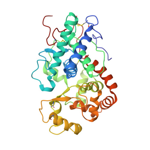
    A cationic class III peroxidase from Sorghum bicolor was purified to homogeneity. The enzyme contains a high-spin heme, as evidenced by UV-visible spectroscopy and EPR. Steady state oxidation of guaiacol was demonstrated and the enzyme was shown to have higher activity in the presence of calcium ions. A Fe(III)/Fe(II) reduction potential of -266 mV vs NHE was determined. Stopped-flow experiments with H2O2 showed formation of a typical peroxidase Compound I species, which converts to Compound II in the presence of calcium. A crystal structure of the enzyme is reported, the first for a sorghum peroxidase. The structure reveals an active site that is analogous to those for other class I heme peroxidase, and a substrate binding site (assigned as arising from binding of indole-3-acetic acid) at the γ-heme edge. Metal binding sites are observed in the structure on the distal (assigned as a Na(+) ion) and proximal (assigned as a Ca(2+)) sides of the heme, which is consistent with the Ca(2+)-dependence of the steady state and pre-steady state kinetics. It is probably the case that the structural integrity (and, thus, the catalytic activity) of the sorghum enzyme is dependent on metal ion incorporation at these positions.


  • Organizational Affiliation
    • Department of Microbiology, University of Nigeria, Nsukka, Nigeria.

Macromolecule Content 

  • Total Structure Weight: 35.64 kDa 
  • Atom Count: 2,807 
  • Modeled Residue Count: 307 
  • Deposited Residue Count: 307 
  • Unique protein chains: 1

Macromolecules

Find similar proteins by:|  3D Structure
Entity ID: 1
MoleculeChains  Sequence LengthOrganismDetailsImage
CATIONIC PEROXIDASE SPC4307Sorghum bicolorMutation(s): 0 
EC: 1.11.1.7
UniProt
Find proteins for P84516 (Sorghum bicolor)
Explore P84516 
Go to UniProtKB:  P84516
Entity Groups
Sequence Clusters30% Identity50% Identity70% Identity90% Identity95% Identity100% Identity
UniProt GroupP84516
Glycosylation
Glycosylation Sites: 2
Sequence Annotations
Expand
Reference Sequence

Oligosaccharides

Help  
Entity ID: 2
MoleculeChains Length2D Diagram GlycosylationD Interactions
alpha-L-fucopyranose-(1-3)-[2-acetamido-2-deoxy-beta-D-glucopyranose-(1-4)]2-acetamido-2-deoxy-beta-D-glucopyranose
B
3N-Glycosylation
Glycosylation Resources
GlyTouCan: G11971MR
GlyCosmos: G11971MR
GlyGen: G11971MR
Entity ID: 3
MoleculeChains Length2D Diagram GlycosylationD Interactions
beta-D-xylopyranose-(1-2)-[alpha-D-mannopyranose-(1-3)]beta-D-mannopyranose-(1-4)-2-acetamido-2-deoxy-beta-D-glucopyranose-(1-4)-[alpha-L-fucopyranose-(1-3)]2-acetamido-2-deoxy-beta-D-glucopyranose
C
6N-Glycosylation
Glycosylation Resources
GlyTouCan: G68461PT
GlyCosmos: G68461PT
GlyGen: G68461PT

Small Molecules

Ligands 5 Unique
IDChains Name / Formula / InChI Key2D Diagram3D Interactions
HEM

Query on HEM


Download:Ideal Coordinates CCD File
D [auth A]PROTOPORPHYRIN IX CONTAINING FE
C34 H32 Fe N4 O4
KABFMIBPWCXCRK-RGGAHWMASA-L
IAC

Query on IAC


Download:Ideal Coordinates CCD File
H [auth A]1H-INDOL-3-YLACETIC ACID
C10 H9 N O2
SEOVTRFCIGRIMH-UHFFFAOYSA-N
GOL

Query on GOL


Download:Ideal Coordinates CCD File
G [auth A]GLYCEROL
C3 H8 O3
PEDCQBHIVMGVHV-UHFFFAOYSA-N
CA

Query on CA


Download:Ideal Coordinates CCD File
E [auth A]CALCIUM ION
Ca
BHPQYMZQTOCNFJ-UHFFFAOYSA-N
NA

Query on NA


Download:Ideal Coordinates CCD File
F [auth A]SODIUM ION
Na
FKNQFGJONOIPTF-UHFFFAOYSA-N

Experimental Data & Validation

Experimental Data

  • Method: X-RAY DIFFRACTION
  • Resolution: 1.27 Å
  • R-Value Free:  0.159 (Depositor), 0.171 (DCC) 
  • R-Value Work:  0.136 (Depositor), 0.148 (DCC) 
  • R-Value Observed: 0.137 (Depositor) 
Space Group: P 31 2 1
Unit Cell:
Length ( Å )Angle ( ˚ )
a = 58.62α = 90
b = 58.62β = 90
c = 208.41γ = 120
Software Package:
Software NamePurpose
REFMACrefinement
XDSdata reduction
Aimlessdata scaling
PHASERphasing

Structure Validation

View Full Validation Report


Ligand Structure Quality Assessment 


Entry History 

Deposition Data

Revision History  (Full details and data files)

  • Version 1.0: 2016-01-13
    Type: Initial release
  • Version 1.1: 2016-03-16
    Changes: Database references
  • Version 2.0: 2020-07-29
    Type: Remediation
    Reason: Carbohydrate remediation
    Changes: Advisory, Atomic model, Data collection, Derived calculations, Other, Structure summary
  • Version 2.1: 2024-01-10
    Changes: Data collection, Database references, Derived calculations, Refinement description, Structure summary
  • Version 2.2: 2024-10-16
    Changes: Structure summary