5ANQ | pdb_00005anq

inhibitors of JumonjiC domain-containing histone demethylases


Experimental Data Snapshot

  • Method: X-RAY DIFFRACTION
  • Resolution: 2.00 Å
  • R-Value Free: 
    0.232 (Depositor), 0.211 (DCC) 
  • R-Value Work: 
    0.189 (Depositor) 
  • R-Value Observed: 
    0.192 (Depositor) 

Starting Model: experimental
View more details

wwPDB Validation 3D Report Full Report

Validation slider image for 5ANQ

Ligand Structure Quality Assessment 


This is version 1.2 of the entry. See complete history

Literature

Substituted 2-(2-Aminopyrimidin-4-Yl)Pyridine-4-Carboxylates as Potent Inhibitors of Jumonjic Domain-Containing Histone Demethylases.

Roatsch, M.Robaa, D.Pippel, M.Nettleship, J.E.Reddivari, Y.Bird, L.E.Hoffmann, I.Franz, H.Owens, R.J.Schole, R.Flaig, R.Sippl, W.Jung, M.

(2016) Future Med Chem 8: 1553

  • DOI: https://doi.org/10.4155/fmc.15.188
  • Primary Citation Related Structures: 
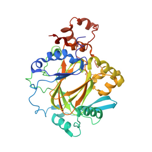
    5ANQ

  • PubMed Abstract: 

    Aberrant expression of iron(II)- and 2-oxoglutarate-dependent JumonjiC histone demethylases has been linked to cancer. Potent demethylase inhibitors are drug candidates and biochemical tools to elucidate the functional impact of demethylase inhibition. Virtual screening identified a novel lead scaffold against JMJD2A with low-micromolar potency in vitro. Analogs were acquired from commercial sources respectively synthesized in feedback with biological testing. Optimized compounds were transformed into cell-permeable prodrugs. A cocrystal x-ray structure revealed the mode of binding of these compounds as competitive to 2-oxoglutarate and confirmed kinetic experiments. Selectivity studies revealed a preference for JMJD2A and JARID1A over JMJD3. Virtual screening and rational structural optimization led to a novel scaffold for highly potent and selective JMJD2A inhibitors.


  • Organizational Affiliation
    • Albert-Ludwigs-University Freiburg, Institute of Pharmaceutical Sciences, Albertstraße 25, 79104 Freiburg i.Br., Germany.

Macromolecule Content 

  • Total Structure Weight: 84.92 kDa 
  • Atom Count: 6,125 
  • Modeled Residue Count: 694 
  • Deposited Residue Count: 718 
  • Unique protein chains: 1

Macromolecules

Find similar proteins by:|  3D Structure
Entity ID: 1
MoleculeChains  Sequence LengthOrganismDetailsImage
LYSINE-SPECIFIC DEMETHYLASE 4A
A, B
359Homo sapiensMutation(s): 0 
EC: 1.14.11.27 (PDB Primary Data), 1.14.11.66 (UniProt), 1.14.11.69 (UniProt)
UniProt & NIH Common Fund Data Resources
Find proteins for O75164 (Homo sapiens)
Explore O75164 
Go to UniProtKB:  O75164
PHAROS:  O75164
GTEx:  ENSG00000066135 
Entity Groups
Sequence Clusters30% Identity50% Identity70% Identity90% Identity95% Identity100% Identity
UniProt GroupO75164
Sequence Annotations
Expand
Reference Sequence

Small Molecules

Ligands 5 Unique
IDChains Name / Formula / InChI Key2D Diagram3D Interactions
5YQ

Query on 5YQ


Download:Ideal Coordinates CCD File
C [auth A],
I [auth B]
2-{2-[(pyridin-3-ylmethyl)amino]pyrimidin-4-yl}pyridine-4-carboxylic acid
C16 H13 N5 O2
YUSWODBVCDCVMS-UHFFFAOYSA-N
SO4

Query on SO4


Download:Ideal Coordinates CCD File
E [auth A],
F [auth A],
K [auth B],
L [auth B]
SULFATE ION
O4 S
QAOWNCQODCNURD-UHFFFAOYSA-L
ZN

Query on ZN


Download:Ideal Coordinates CCD File
G [auth A],
M [auth B]
ZINC ION
Zn
PTFCDOFLOPIGGS-UHFFFAOYSA-N
FE2

Query on FE2


Download:Ideal Coordinates CCD File
D [auth A],
J [auth B]
FE (II) ION
Fe
CWYNVVGOOAEACU-UHFFFAOYSA-N
CL

Query on CL


Download:Ideal Coordinates CCD File
H [auth A],
N [auth B],
O [auth B],
P [auth B]
CHLORIDE ION
Cl
VEXZGXHMUGYJMC-UHFFFAOYSA-M

Experimental Data & Validation

Experimental Data

  • Method: X-RAY DIFFRACTION
  • Resolution: 2.00 Å
  • R-Value Free:  0.232 (Depositor), 0.211 (DCC) 
  • R-Value Work:  0.189 (Depositor) 
  • R-Value Observed: 0.192 (Depositor) 
Space Group: P 21 21 2
Unit Cell:
Length ( Å )Angle ( ˚ )
a = 100.694α = 90
b = 149.271β = 90
c = 57.527γ = 90
Software Package:
Software NamePurpose
REFMACrefinement
XDSdata reduction
Aimlessdata scaling
MOLREPphasing

Structure Validation

View Full Validation Report


Ligand Structure Quality Assessment 


Entry History 

Revision History  (Full details and data files)

  • Version 1.0: 2016-03-23
    Type: Initial release
  • Version 1.1: 2016-09-28
    Changes: Database references
  • Version 1.2: 2024-01-10
    Changes: Data collection, Database references, Derived calculations, Other, Refinement description