5AFW | pdb_00005afw

Assembly of methylated LSD1 and CHD1 drives AR-dependent transcription and translocation


Experimental Data Snapshot

  • Method: X-RAY DIFFRACTION
  • Resolution: 1.60 Å
  • R-Value Free: 
    0.227 (Depositor), 0.234 (DCC) 
  • R-Value Work: 
    0.209 (Depositor), 0.214 (DCC) 
  • R-Value Observed: 
    0.210 (Depositor) 

Starting Model: experimental
View more details

wwPDB Validation 3D Report Full Report

Validation slider image for 5AFW

This is version 1.3 of the entry. See complete history

Literature

Assembly of Methylated Kdm1A and Chd1 Drives Androgen Receptor-Dependent Transcription and Translocation.

Metzger, E.Willmann, D.Mcmillan, J.Forne, I.Metzger, P.Gerhardt, S.Petroll, K.Von Maessenhausen, A.Urban, S.Schott, A.Espejo, A.Eberlin, A.Wohlwend, D.Schule, K.M.Schleicher, M.Perner, S.Bedford, M.T.Jung, M.Dengjel, J.Flaig, R.Imhof, A.Einsle, O.Schule, R.

(2016) Nat Struct Mol Biol 23: 132

  • DOI: https://doi.org/10.1038/nsmb.3153
  • Primary Citation Related Structures: 
    5AFW

  • PubMed Abstract: 

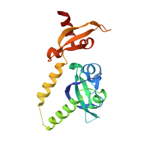
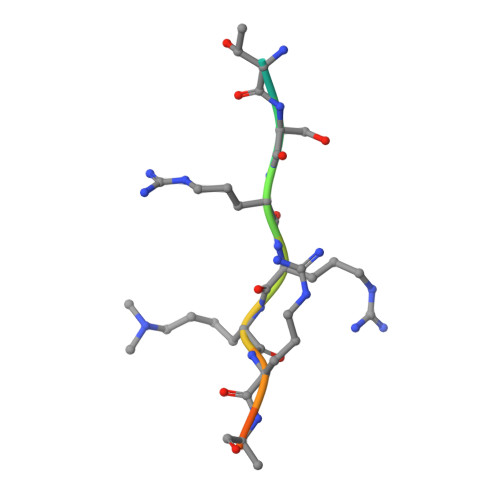
    Prostate cancer evolution is driven by a combination of epigenetic and genetic alterations such as coordinated chromosomal rearrangements, termed chromoplexy. TMPRSS2-ERG gene fusions found in human prostate tumors are a hallmark of chromoplexy. TMPRSS2-ERG fusions have been linked to androgen signaling and depend on androgen receptor (AR)-coupled gene transcription. Here, we show that dimethylation of KDM1A at K114 (to form K114me2) by the histone methyltransferase EHMT2 is a key event controlling androgen-dependent gene transcription and TMPRSS2-ERG fusion. We identified CHD1 as a KDM1A K114me2 reader and characterized the KDM1A K114me2-CHD1 recognition mode by solving the cocrystal structure. Genome-wide analyses revealed chromatin colocalization of KDM1A K114me2, CHD1 and AR in prostate tumor cells. Together, our data link the assembly of methylated KDM1A and CHD1 with AR-dependent transcription and genomic translocations, thereby providing mechanistic insight into the formation of TMPRSS2-ERG gene fusions during prostate-tumor evolution.


  • Organizational Affiliation
    • Klinik für Urologie und Zentrale Klinische Forschung, Klinikum der Universität Freiburg, Freiburg, Germany.

Macromolecule Content 

  • Total Structure Weight: 22.73 kDa 
  • Atom Count: 1,579 
  • Modeled Residue Count: 170 
  • Deposited Residue Count: 186 
  • Unique protein chains: 2

Macromolecules

Find similar proteins by:|  3D Structure
Entity ID: 1
MoleculeChains  Sequence LengthOrganismDetailsImage
CHROMODOMAIN-HELICASE-DNA-BINDING PROTEIN 1174Homo sapiensMutation(s): 0 
EC: 3.6.4.12 (PDB Primary Data), 3.6.4 (UniProt)
UniProt & NIH Common Fund Data Resources
Find proteins for O14646 (Homo sapiens)
Explore O14646 
Go to UniProtKB:  O14646
PHAROS:  O14646
GTEx:  ENSG00000153922 
Entity Groups
Sequence Clusters30% Identity50% Identity70% Identity90% Identity95% Identity100% Identity
UniProt GroupO14646
Sequence Annotations
Expand
Reference Sequence
Find similar proteins by:  Sequence   |   3D Structure  
Entity ID: 2
MoleculeChains  Sequence LengthOrganismDetailsImage
LYSINE-SPECIFIC HISTONE DEMETHYLASE 1A12Homo sapiensMutation(s): 0 
EC: 1 (PDB Primary Data), 1.14.11 (UniProt), 1.14.99.66 (UniProt), 1.14.11.65 (UniProt)
UniProt & NIH Common Fund Data Resources
Find proteins for O60341 (Homo sapiens)
Explore O60341 
Go to UniProtKB:  O60341
PHAROS:  O60341
GTEx:  ENSG00000004487 
Entity Groups
Sequence Clusters30% Identity50% Identity70% Identity90% Identity95% Identity100% Identity
UniProt GroupO60341
Sequence Annotations
Expand
Reference Sequence

Small Molecules

Ligands 2 Unique
IDChains Name / Formula / InChI Key2D Diagram3D Interactions
EDO

Query on EDO


Download:Ideal Coordinates CCD File
C [auth A]
D [auth A]
E [auth A]
F [auth A]
G [auth A]
C [auth A],
D [auth A],
E [auth A],
F [auth A],
G [auth A],
H [auth A],
I [auth A],
J [auth A],
K [auth A]
1,2-ETHANEDIOL
C2 H6 O2
LYCAIKOWRPUZTN-UHFFFAOYSA-N
CL

Query on CL


Download:Ideal Coordinates CCD File
L [auth A],
M [auth A],
N [auth A]
CHLORIDE ION
Cl
VEXZGXHMUGYJMC-UHFFFAOYSA-M
Modified Residues  1 Unique
IDChains TypeFormula2D DiagramParent
MLY
Query on MLY
B
L-PEPTIDE LINKINGC8 H18 N2 O2LYS

Experimental Data & Validation

Experimental Data

  • Method: X-RAY DIFFRACTION
  • Resolution: 1.60 Å
  • R-Value Free:  0.227 (Depositor), 0.234 (DCC) 
  • R-Value Work:  0.209 (Depositor), 0.214 (DCC) 
  • R-Value Observed: 0.210 (Depositor) 
Space Group: P 21 21 2
Unit Cell:
Length ( Å )Angle ( ˚ )
a = 110.296α = 90
b = 44.445β = 90
c = 46.194γ = 90
Software Package:
Software NamePurpose
BUSTERrefinement
XDSdata reduction
XDSdata scaling
PHASERphasing

Structure Validation

View Full Validation Report


Entry History 

Revision History  (Full details and data files)

  • Version 1.0: 2016-01-13
    Type: Initial release
  • Version 1.1: 2016-01-20
    Changes: Database references
  • Version 1.2: 2016-03-02
    Changes: Database references
  • Version 1.3: 2024-01-10
    Changes: Data collection, Database references, Derived calculations, Other, Refinement description