5AFG | pdb_00005afg

Structure of the Stapled Peptide Bound to Mdm2


Experimental Data Snapshot

  • Method: X-RAY DIFFRACTION
  • Resolution: 1.90 Å
  • R-Value Free: 
    0.226 (Depositor), 0.235 (DCC) 
  • R-Value Work: 
    0.176 (Depositor), 0.189 (DCC) 
  • R-Value Observed: 
    0.179 (Depositor) 

Starting Model: experimental
View more details

wwPDB Validation 3D Report Full Report

Validation slider image for 5AFG

Ligand Structure Quality Assessment 


This is version 2.1 of the entry. See complete history

Literature

Double Strain-Promoted Macrocyclization for the Rapid Selection of Cell-Active Stapled Peptides.

Lau, Y.H.Wu, Y.Rossmann, M.Tan, B.X.De Andrade, P.Tan, Y.S.Verma, C.Mckenzie, G.J.Venkitaraman, A.R.Hyvonen, M.Spring, D.R.

(2015) Angew Chem Int Ed Engl 54: 15410

  • DOI: https://doi.org/10.1002/anie.201508416
  • Primary Citation Related Structures: 
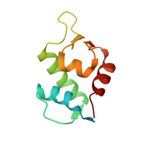
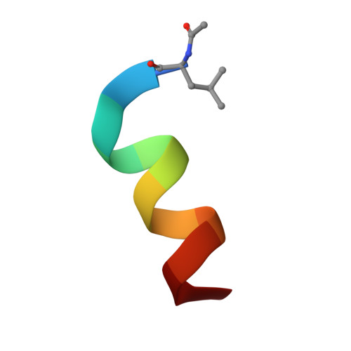
    5AFG

  • PubMed Abstract: 

    Peptide stapling is a method for designing macrocyclic alpha-helical inhibitors of protein-protein interactions. However, obtaining a cell-active inhibitor can require significant optimization. We report a novel stapling technique based on a double strain-promoted azide-alkyne reaction, and exploit its biocompatibility to accelerate the discovery of cell-active stapled peptides. As a proof of concept, MDM2-binding peptides were stapled in parallel, directly in cell culture medium in 96-well plates, and simultaneously evaluated in a p53 reporter assay. This in situ stapling/screening process gave an optimal candidate that showed improved proteolytic stability and nanomolar binding to MDM2 in subsequent biophysical assays. α-Helicity was confirmed by a crystal structure of the MDM2-peptide complex. This work introduces in situ stapling as a versatile biocompatible technique with many other potential high-throughput biological applications.


  • Organizational Affiliation
    • Department of Chemistry, University of Cambridge, Lensfield Rd, Cambridge CB2 1EW (UK).

Macromolecule Content 

  • Total Structure Weight: 12.87 kDa 
  • Atom Count: 913 
  • Modeled Residue Count: 95 
  • Deposited Residue Count: 109 
  • Unique protein chains: 2

Macromolecules

Find similar proteins by:|  3D Structure
Entity ID: 1
MoleculeChains  Sequence LengthOrganismDetailsImage
E3 UBIQUITIN-PROTEIN LIGASE MDM297Homo sapiensMutation(s): 2 
EC: 6.3.2.19 (PDB Primary Data), 2.3.2.27 (UniProt)
UniProt & NIH Common Fund Data Resources
Find proteins for Q00987 (Homo sapiens)
Explore Q00987 
Go to UniProtKB:  Q00987
PHAROS:  Q00987
GTEx:  ENSG00000135679 
Entity Groups
Sequence Clusters30% Identity50% Identity70% Identity90% Identity95% Identity100% Identity
UniProt GroupQ00987
Sequence Annotations
Expand
Reference Sequence
Find similar proteins by:  Sequence   |   3D Structure  
Entity ID: 2
MoleculeChains  Sequence LengthOrganismDetailsImage
STAPLED PEPTIDE12synthetic constructMutation(s): 0 

Small Molecules

Ligands 1 Unique
IDChains Name / Formula / InChI Key2D Diagram3D Interactions
P07

Query on P07


Download:Ideal Coordinates CCD File
C [auth B]1,8-DIETHYL-1,8-DIHYDRODIBENZO[3,4:7,8][1,2,3]TRIAZOLO[4',5':5,6]CYCLOOCTA[1,2-D][1,2,3]TRIAZOLE
C20 H18 N6
YRZRZRBFIALCLI-MKVCYXFSSA-N
Modified Residues  1 Unique
IDChains TypeFormula2D DiagramParent
LAY
Query on LAY
B
L-PEPTIDE LINKINGC8 H15 N O3LEU

Experimental Data & Validation

Experimental Data

  • Method: X-RAY DIFFRACTION
  • Resolution: 1.90 Å
  • R-Value Free:  0.226 (Depositor), 0.235 (DCC) 
  • R-Value Work:  0.176 (Depositor), 0.189 (DCC) 
  • R-Value Observed: 0.179 (Depositor) 
Space Group: P 41 21 2
Unit Cell:
Length ( Å )Angle ( ˚ )
a = 36.821α = 90
b = 36.821β = 90
c = 177.416γ = 90
Software Package:
Software NamePurpose
REFMACrefinement
PROTEUM2data reduction
PROTEUM2data scaling
PHASERphasing

Structure Validation

View Full Validation Report


Ligand Structure Quality Assessment 


Entry History 

Deposition Data

Revision History  (Full details and data files)

  • Version 1.0: 2016-01-27
    Type: Initial release
  • Version 2.0: 2019-05-15
    Changes: Data collection, Derived calculations, Other, Polymer sequence
  • Version 2.1: 2024-01-10
    Changes: Data collection, Database references, Derived calculations, Other, Refinement description